-
PDF
- Split View
-
Views
-
Cite
Cite
Metasebia W Abebe, Mary Mesfin Nigussie, Retroperitoneal necrotizing fasciitis with the involvement of the anterior abdominal wall following perianal abscess, Journal of Surgical Case Reports, Volume 2021, Issue 6, June 2021, rjab222, https://doi.org/10.1093/jscr/rjab222
Close - Share Icon Share
Abstract
Necrotizing fasciitis (NF) is a life-threatening infection, which requires immediate debridement and broad-spectrum antibiotic treatment. Delay in prompt diagnosis and operative debridement is associated with significant morbidity and mortality. Retroperitoneal NF is a rare condition whereby the infection within the pelvis or retroperitoneum rapidly expands over the fascial planes to involve the anterior abdominal wall (AW), the thighs and the buttocks. It presents a challenge for surgical access due to the anatomic depth of the structures and may result in extensive soft tissue loss requiring complex AW reconstruction for closure. The case discussed here is a 43-year-old female with a perianal abscess that progressed to retroperitoneal and anterior AW NF with intra-peritoneal abscess collection requiring bilateral tensor fascia-lata graft for the closure of the anterior AW fascia defect.
INTRODUCTION
Necrotizing fasciitis (NF) is a life-threatening infection and widespread necrosis of the subcutaneous tissue and fascia often accompanied by systemic inflammatory response syndrome and significant mortality necessitating immediate resuscitation, surgical intervention with debridement of dead tissue and broad-spectrum antibiotic treatment [1–5].
Depending on type and number of causative organisms, four types of NF have been described [4, 6–8]: Type 1—accounting for the majority of cases and having a polymicrobial origin, Type 2—being monomicrobial, Type 3—gram-negative and often marine-related, whereas Type 4 is of fungal etiology. Both aerobic and anaerobic bacteria alone or in synergism and fungal species have been identified as the causative agents in NF [4, 7]. Although NF can occur in the absence of clinical risk factors [5, 6], older age, diabetes mellitus, obesity, cancers and autoimmune disorders are identified clinical risk factors [2–5].
The clinical feature common to all types of NF is agonizing pain, quite out of proportion with few local clinical signs [2, 7]. In the absence of an early diagnosis, resuscitation and surgical management, mortality from NF ranges from 8.7 to 73% [2, 3, 9] with recent literature reporting a lesser rate of mortality around 26-32% [4, 5].
Common sites of infection are the lower extremities, abdomen and perineum with [3, 4, 6, 7] perianal conditions as abscess rarely resulting in potentially fatal complications as retroperitoneal NF [2, 3, 10].
Retroperitoneal NF is a rare condition [2, 10], where the infection invades into the pelvis or retroperitoneum from the more superficial fascial levels along interfascial planes, where it makes surgical management even more difficult because of the deep space within the pelvis and retro-peritoneum [2, 3].
A high index of suspicion is necessary for retroperitoneal NF as any recent history of a perianal abscess, surgery or any trauma to the skin can be the inciting event leading to an inoculum of bacteria into the host’s system [2, 3]. In addition to the initial prompt diagnosis, resuscitation and surgical debridement, re-look surgery should be considered after 24–48 hours to reassess and re-debride if indicated. The absence of progression of the infective process is a good prognostic sign and can be used to plan further management, whereas untreated or inadequately debrided NF may rapidly progress into septic shock with mortality rate close to 100% [2, 3, 10].
Retroperitoneal NF may require leaving the abdomen open with a temporary abdominal closure device [3]. Following thorough debridement of dead tissue and re-look surgery, simple to complex reconstruction methods may be needed to recreate the native abdominal wall (AW) [4]. The goals of the reconstructive surgery in the management of complex AW defects are to restore the structural and functional continuity of the muscle-fascial system, provide stable coverage and achieve local wound closure [4].
CASE REPORT
A 43-year-old female presented with continuing fever, lower abdominal pain and foul-smelling discharge from her left perianal region of 2 weeks duration. She had no previous medical illness. Upon physical examination of the perineum, necrotic tissue, foul-smelling thin pus were observed.
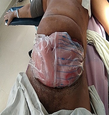
The patient was taken in for surgery—the left perianal region was debrided revealing deep pockets of abscess. By the second postoperative day, lower abdominal pain worsened and ultrasound showed a fluid collection. Laparotomy revealed thick abscess in the entire anterior AW and the rectus sheath with necrotic rectus muscle, superiorly extending to the subcostal region and inferiorly to the retro-pubic space. The abscess was also in the peritoneal cavity, laterally extending to the para-renal retroperitoneal region. Abscess drainage and debridement done and abdomen left open with Bogota bag (Fig. 1) with pockets packed and drainage tubes in situ.

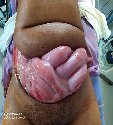
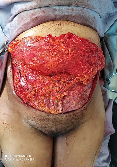
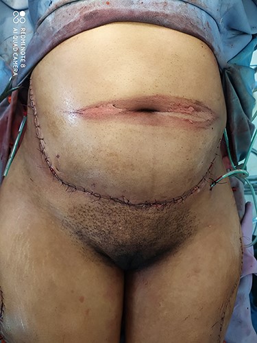
The patient was transferred to intensive care unit (ICU) postoperatively, antibiotics changed to a wider spectrum of coverage. Relaparotomy was done 48 hours later but showed no disease progression. The patient stayed in ICU and continued antibiotics for 2 weeks. On the 35th day following initial debridement, 15 × 30 cm sub-umbilical fascia defect (Fig. 2) was reconstructed with non-vascularized bilateral tensor fascia-lata graft (Fig. 3) and abdomen was closed (Fig. 4). Secondary closure of the perianal wound was also done. At 3, 6 and 9-month follow-up visits, the patient is doing well and the computed tomography scan shows normal anterior abdominal fascia.

Open abdominal wound with 15 × 30 cm sub-umbilical fascia defect.

Fascial closure with non vascularized bilateral tensor fascia lata graft.

DISCUSSION
NF is a rare but rapidly spreading fulminant soft tissue infection wherein the absence of timely intervention may results in complications as septic shock and multi-organ failure and high mortality [6–8, 10]. A period of 2 weeks lapsed before the initial surgical intervention that probably contributed to the spread of the infection in our patient— a 43-year-old female with no identifiable risk factors.
Even with adequate care, patients frequently suffer substantial morbidity and require reconstruction and rehabilitation [8]. Our patient also had significant morbidity that required 2 weeks of ICU admission and care, prolonged admission to the surgical ward and several reoperations to control the infection and finally reconstruct the AW.
The mean duration of antibiotic therapy described for NF is 4–6 weeks [6], which was also the case in our patient where a perianal abscess progressed to pelvic abscess with NF of retro-peritoneum and anterior AW fascia that finally necessitated a complex abdominal reconstruction with non-vascularized tensor fascia graft since one of the structures forming the scaffolding for intra-abdominal contents—the fascia was completely debrided in the process of controlling the infection.
Reconstruction of the AW focuses primarily on the restoration of function and stability, prevention of viscera herniation, as well as the re-establishment of esthetically acceptable appearance. The AW defects are divided into two groups depending on the presence or absence of normal skin coverage. In Type I defect, there is intact or stable covering skin, whereas Type II defects have absent or unstable skin cover [2]. In the absence of microvascular free flap reconstruction or vacuum-assisted closure therapy for fast and effective wound closure, we have used a free tensor fascia graft for reconstructing the fascial defect of Type I anterior AW defect in our patient.
CONFLICT OF INTEREST STATEMENT
Authors have no conflict of interest to declare.
References
- antibiotics
- debridement
- abscess
- buttocks
- fascia lata
- necrotizing fasciitis
- reconstructive surgical procedures
- retroperitoneal space
- surgical procedures, operative
- thigh
- tissue transplants
- infections
- diagnosis
- fascia
- morbidity
- mortality
- pelvis
- peritoneum
- perianal abscess
- abdominal wall, anterior
- soft tissue


