-
PDF
- Split View
-
Views
-
Cite
Cite
Ali Al Ansari, Shehab Ahmed, Eyad Mansour, Maher A Abass, Idiopathic myointimal hyperplasia of the mesenteric veins, Journal of Surgical Case Reports, Volume 2021, Issue 1, January 2021, rjaa453, https://doi.org/10.1093/jscr/rjaa453
Close - Share Icon Share
Abstract
Idiopathic myointimal hyperplasia of the mesenteric veins (IMHMV) is caused by proliferation of smooth muscle cells in the wall of small mesenteric veins and venules with accumulation of a proteoglycan matrix leading to a non-thrombotic, non-inflammatory venous occlusion resulting in venous ischemia. IMHMV is a rare and poorly understood disease, with <20 case reports in the literature. The purpose of this report is to describe the case of a 63-year-old man who presented with this condition that resulted in colonic ischemia necessitating surgical resection. The cause of IMHMV in this patient was attributed to a Chinese herbal supplement used for degenerative osteoarthritis of the knees. A brief review of the literature is provided along with the case report.
INTRODUCTION
Non-thrombotic, non-inflammatory occlusion of the mesenteric veins is a rare phenomenon and can be classified into three distinct yet closely related pathologies; mesenteric inflammatory veno-occlusive disease (MIVOD), idiopathic mesenteric phlebosclerosis (IMP) and idiopathic myointimal hyperplasia of the mesenteric veins (IMHMV). Their presentations are similar to inflammatory bowel disease (IBD) and this proves to be a diagnostic challenge for physicians. It has already been discovered that Chinese herbal remedies is a causative factor in IMP; however, it has not been fully elucidated in IMHMV. This case hypothesizes the link between Chinese herbal remedies causing IMHMV.

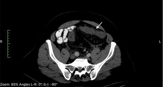
CT of the abdomen at time of admission reveals thickening of the sigmoid colon (white arrows).
![Repeat computed tomography of the abdomen 12 days after admission reveals progressive thickening of the sigmoid colon [long white arrow] and free fluid [short white arrow].](https://oupdevcdn.silverchair-staging.com/oup/backfile/Content_public/Journal/jscr/2021/1/10.1093_jscr_rjaa453/3/m_rjaa453f2.jpeg?Expires=1773987937&Signature=eaW3JL57wjA4ZGMqf05AcyDNyIbyON-ZoEWCMQOobwA5bGUcxzTeJGybhWB1TNBJuGYz4Z~IcFiKwvyPxqqgPxhLsyTypZKkb3X6qic62WI1sq5gyG-JEitA8RNnIt0EynrYlEHq2J5lfpoQ5PBtuhwPzCM0sGgy0EBxGf2dess5Z-InzdUQ9G~dlw2-vO4ktYRoPBiD7e8H36NpX54rCabrF80FsUseRZuufQVQCJw3GAFW2fLEI57mIlz6K-YfgZkH3BFNVDJrvbn2SbJ7LVgXmRKzChIrqTpEIDyXKaVpC8larV-tHx29GqM9fSl6M3GCHV8jYT9dp2I71GA7vQ__&Key-Pair-Id=APKAIYYTVHKX7JZB5EAA)
Repeat computed tomography of the abdomen 12 days after admission reveals progressive thickening of the sigmoid colon [long white arrow] and free fluid [short white arrow].
CASE REPORT
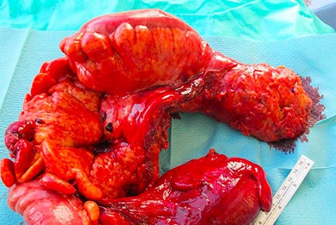
A 63-year-old Middle-Eastern man developed sudden onset of lower abdominal pain and non-bloody diarrhea. At time of presentation there was no report of fever, chills, nausea or emesis. The patient was overall healthy except for a history of benign prostatic hyperplasia and degenerative osteoarthritis of the knees, which he treated for several months with a Chinese herbal supplementation. The past surgical history was significant for tonsillectomy and inguinal hernia repair. The patient had received a course of oral ciprofloxacin due to worsening of his diarrhea. He was admitted to the hospital and laboratory results were significant for an elevated C-reactive protein at 140 mg/L. Computed tomography (CT) scan revealed left sided colitis involving the sigmoid and descending colon (Fig. 1). Serosal irregularity and pericolic inflammation change with mesocolic vascular congestion and hyperemia. Colonoscopy showed edema and erythema without gross ulceration. An additional course of intravenous ciprofloxacin and metronidazole was unsuccessful in resolving the symptoms leading to the administration of ertapenem and azithromycin by the treating gastroenterologist. Subsequent stool studies were positive for Entamoeba histolytica prompting a full course of oral tinidazole. With worsening of the patient’s symptoms, a second CT scan performed 12 days later demonstrated interval progression of the colitis to the distal transverse colon with free intra-abdominal fluids (Fig. 2). Repeat colonoscopy to assess for inflammatory bowel disease showed diffuse edema and erythema without deep ulceration. Biopsies revealed non-specific severe colitis. Despite bowel rest with total parenteral nutrition and the various intravenous and oral antibiotics, the patient continued to deteriorate prompting an extended left hemicolectomy with end transverse colostomy and a low Hartmann’s rectal pouch 26 days after initial presentation. Examination of the 75 cm specimen revealed macroscopic features of ischemia with indurated brown-reddish bowel wall and bulky hardened mesenteric fat tissue (Fig. 3). Gross inspection of the inflamed mucosa revealed a fibrinous layer. Histopathologic assessment showed inflammation and fibrosis of the mucosa, especially of the lamina propria with rarefaction of the crypts. Proliferation of small vessels was visible in the lamina propria, the submucosa and the pericolic fat. Moreover, some vessels showed a fibromyxoid wall-thickening. Elastic van Gieson (EvG) stains revealed massive changes in venous structures with hyperplasia of the cellular and matrical mass in the initmal layer leading to subtotal occlusion of the venous lumens. Histology identified focal secondary thrombosis. Some lymph nodes were rich of plasma cells with a dilated sinus with ectasia of the lymph vessels. The findings were consistent with IMHMV.

Examination of the 75 cm specimen revealed macroscopic features of ischemia with indurated brown-reddish bowel wall and bulky hardened mesenteric fat tissue.
The postoperative course was significant for a prolonged recovery complicated by intraabdominal fluid collections requiring intravenous antibiotics and a CT-guided drainage. After 25 days of the operative intervention (66 days from onset of symptoms), the patient was discharged home, and 5 months later he underwent reversal of stoma with transversorectostomy. After 5 years, the patient continues to do well and surveillance colonoscopy shows normal colon and anastomosis.
DISCUSSION
IMHMV is a rare and poorly understood disease with unknown etiology, characterized by smooth muscle proliferation leading to non-thrombotic, non-inflammatory mesenteric ischemia. Patients with IMHMV present with overlapping symptoms with IBD. With <20 reported cases in the literature, IMHMV remains a diagnostic challenge for physicians [1]. Difficulties in recognizing IMHMV are 2-fold. It presents with a sudden onset of lower abdominal cramps and diarrhea that progress to a colicky pain with weight loss often leading to a clinical misdiagnosis of IBD. Secondly, its radiological findings are somewhat similar with non-specific ulcerated lesions in the left colon favoring the sigmoid. However, some cases have been reported to involve the entire colon and terminal ileum. Key differences between IBD and IMHMV are best appreciated in the histopathologic evaluation. IMHMV features microscopic non-inflamed, non-thrombosed thickened venules with hypertrophy of the smooth muscle in the intimal layer causing reduced perfusion and ischemia. However, the diagnosis is difficult to make based on superficial endoscopic biopsies and is more evident when a portion of the colon is submitted for full thickness evaluation.
IMHMV was first reported in 1991 and nearly 3 decades later there is a paucity of available studies to shed light on the etiology and pathogenesis of this disorder [2]. Abu Alfa et al. [3] noted a striking resemblance of veins in IMHMV and those of patients with failed coronary artery bypass venous grafts. As such the hypothesis of ‘arterialization’ in the venous system as a contributing factor to the development of veno-occlusive disease in the mesentery has been proposed. Flaherty et al. [4] described seven patients with what they termed ‘mesenteric inflammatory veno-occlusive disease’ (MIVOD), another rare cause of intestinal ischemia highlighted by the presence of lymphocytic inflammatory infiltrate. Interestingly enough myointimal hyperplasia in the mesenteric veins was identified in three of the seven patients. This finding led them to the hypothesis that MIVOD may in fact be a precursor to IMHMV; however, confirmation of this relation has yet to be confirmed. Mesenteric veno-occlusive disease in the absence of a hypercoagulable state, sepsis, trauma or pancreatitis is uncommon and covers the spectrum of IMHMV, MIVOD and idiopathic mesenteric phlebosclerosis (IMP). Diagnosis of these diseases requires biopsy and microscopic sampling as each have differentiating characteristics and respond differently to medical management. In the case of IMP, Chinese herbal medications have been implicated as a causative factor and most cases of IMP have been reported in East Asia [5, 6]. A Chinese herbal medication for knee osteoarthritis was the only medication in our case that could have explained the development of IMHMV and it was initiated 2 months prior to presentation. We hypothesize that the medication which typically stimulates cartilage growth might have triggered the smooth muscle layer proliferation in the mesenteric veins.
CONFLICT OF INTEREST STATEMENT
None declared.


