-
PDF
- Split View
-
Views
-
Cite
Cite
Jay Lodhia, David Msuya, Rune Philemon, Adnan Sadiq, Alex Mremi, Gastric perforation resulting into pneumoperitoneum in a neonate: a case report, Journal of Surgical Case Reports, Volume 2020, Issue 8, August 2020, rjaa298, https://doi.org/10.1093/jscr/rjaa298
Close - Share Icon Share
Abstract
Gastric perforation in a neonate is a rare surgical emergency in routine practice. The causes and predisposing factors for gastric perforation in a neonate vary from traumatic to benign conditions like inflammatory processes. Early detection, intensive care, stabilization and prompt surgery yield positive outcome. Early diagnosis is important for better prognosis. Simple investigation such as plain abdominal X-ray can adequately lead to the diagnosis by showing pneumoperinoneum. We present a 3-day-old neonate; born at term who presented with abdominal distension and vomiting. Plain abdominal X-ray revealed pneumoperitoneum. Emergency laparotomy was performed where a gastric perforation was found measuring 0.5 by 0.5 cm located on the anterior aspect of the stomach body near the pylorus. The baby underwent successful surgical intervention and recovered well.
INTRODUCTION
Gastric perforation in neonates is a rare surgical emergency [1]. It is said that it accounts for 7% of all gastrointestinal perforations in neonates with poor outcomes but early diagnosis and surgical treatment increases survival rates [1–4]. Neonates with peritonitis due to gastric perforation can present with features of intestinal obstruction. The exact causes of gastric perforation remains unclear but factors can either be spontaneous like in preterm babies and secondary from obstruction or trauma [2]. Our report is of a case of a full-term newborn with a single spontaneous perforation located on the anterior aspect of the stomach body near the pylorus, which was surgically treated.
CASE REPORT
A 3-day-old female baby was admitted to our center due to a 2-day history of postprandial vomiting of the ingested breast milk, which was of non-projectile. It was also associated with passage of black stools that were ‘sticky’ in nature as reported by the mother, low-grade fever which responded to paracetamol suspension and gradual abdominal distension. The baby was born normally via spontaneous vaginal delivery at term with a birth weight of 2.5 kg. The mother reported normal pregnancy.
Upon examination, an active baby with hemodynamic stable vitals. The abdomen was symmetrically distended with visible superficial blood veins, tender to palpate all over and reduced bowel sounds on auscultation. The respiratory, cardiovascular, genitourinary and nervous systems examination was essentially normal.
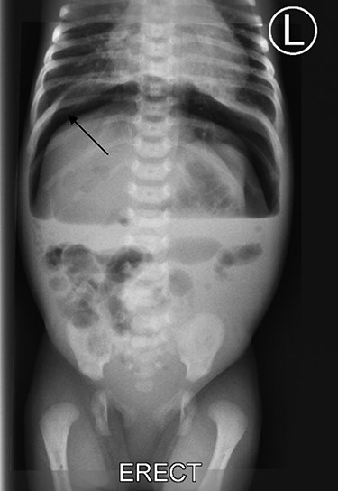
Provisional diagnosis of intestinal obstruction due to necrotizing enterocolitis with early neonatal sepsis was made. Initial blood investigations showed hemoglobin of 15.4 g/dl, BUN of 1.72 mmol/l and a serum creatinine of 72 μmol/l. Abdominal-pelvic ultrasound w revealed focal dilated bowel loops with poor peristalsis, free intraabdominal fluid. The liver, spleen and kidneys had normal echo-texture and impression of pneumoperitoneum was made. Plain abdominal X-ray showed features of pneumoperitoneum (Fig. 1).

Plain abdominal X-ray shows free air under right dome of diaphragm (arrow).
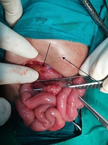
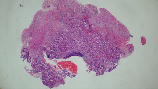
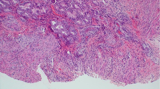
The baby was prepared and taken for an emergency laparotomy whereby intraoperatively, a gastric perforation was found measuring 0.5 by 0.5 cm, circumferential and located on the anterior aspect of the stomach body near the pylorus (Fig. 2). There was also about 50 ml of amber-colored ascites. The perforation was repaired and Grahm’s patch was put, thorough abdominal lavage and the abdomen was closed in layers. Biopsy from the perforation site revealed non-necrotizing mild chronic gastritis, not otherwise specified with ulcerations (Figs 3 and 4).

Photograph showing gastric perforation and abdominal distension (arrows).

Histopathology of the biopsy from the gastric perforation showing near full thickness with non-specific mixed chronic inflammation (H&E staining, x10).

Histological appearance of gastric wall showing ulceration, mild chronic inflammation, focal hemorrhage and serosal fibrosis (H&E staining, ×20).
Postoperatively the baby faired well, started oral feeds on day-2 and tolerated. The baby developed mild surgical site infection on day 6 that was treated with daily dressing and systemic antibiotics and discharged on day 28.
DISCUSSION
Neonatal gastric perforation is a rare surgical emergency with an incidence of 1:2900 live births [1]. Causes of perforation can be spontaneous (idiopathic) or secondary. Secondary causes include iatrogenic injury from nasogastric tubes, mechanical ventilation or mechanical (or functional) gastric obstructions from atresia, hypertrophic pyloric stenosis, meconium ileus or webs [1, 2, 4]. Others causes include meconium gastritis, Meckel’s diverticulum and intestinal malrotation [1, 2]. Idiopathic cause includes preterm neonates due to the lack of gastric maturity and protective factors, such as lack of C-KIT mast cells and intestinal pacemaker cells [6]. This can be augmented with other factors, such as stress, asphyxia and small for gestational age [1, 4, 9]. None of these factors were associated with our case as the baby was born at term, appropriate for gestational age. As reported histologically, the biopsy from perforation site indicated non-specific gastritis with ulceration. Nevertheless it is said that irrespective of the etiology, most neonatal gastric perforation occurs between 2 and 7 days of life [4, 8]. The overall survival rate has improved overtime due to the advancement in the intensive care units although mortality rates have been reported ~70% [6].
A study by Farrugia et al. outlined that gastrointestinal perforation occurs as a complication from necrotizing enterocolitis in 42% of the cases with a mortality rate of 62%. The authors stated that mortality rate was 14% [5]. From the index case, presentation of necrotizing enterocolitis was not found intraoperatively or histologically.
Another case report by Obeida et al. revealed a rather rare cause of gastric perforation. The authors reported of a term baby with congenital diaphragmatic hernia where a perforation was found intraoperatively on the stomach presumably due to necrosis. The perforation was repaired surgically with Vicryl absorbable suture, and the hernia defect repaired too [7]. In our case, the stomach looked innocent but the perforation was primarily repaired with absorbable suture similarly reported by the authors.
A wide range of clinical features can be presented in neonates, ranging from signs of septicemia to intestinal obstruction due to paralytic ileus from peritonitis from contamination [3, 8]. The authors diagnosed the pneumoperinoneum from a plain abdominal X-ray showing free gas under the right hemi-diaphragm as this was also the case in the index case. Other modalities can be considered like contrast studies where a leakage of the contrast can be seen into the peritoneum cavity which can aid in the diagnosis and point the site of perforation in the gastrointestinal tract [5].
Surgery remains the mainstay as the definitive management [9]. Gastrorrhaphy is the procedure of choice and rarely is gastrectomy to be performed [6, 9]. In addition, underlying anatomical cause should be identified and corrected [2, 6]. Early detection of the diagnosis meanwhile stabilization of the patient is vital in the outcome. Intensive care is ideal where the neonate is monitored closely, electrolyte and hydration balance is maintained and respiratory assistance may be required [1]. Protective nasogastric tube can be retained postsurgery to avoid gastric filling and distension hence protecting the repaired site [1, 2]. Poor prognostic factors include male gender, low birth weight, metabolic acidosis, hyponatremia and time between symptoms and surgical intervention [2, 3, 4, 9]. The index case did not have any of the mentioned poor prognostic factors and surgery was performed promptly.
CONCLUSION
Neonatal gastric perforation is a rare life threatening surgical emergency. Prematurity is the most common associated risk factor. High suspicion of index based on clinical judgment due to unspecific clinical signs should be augmented by prompt investigations is essential for diagnosis. Early surgical management can improve survival with the aid of quality intensive care and follow-up.
Authors’ contributions
J.L. conceptualized and the prepared the initial draft of the manuscript. D.M. and J.L. performed the operation. A.S. prepared and reported the radiological films. A.M. reported prepared the histology images and prepared the final version of the manuscript. R.P. provided the technical input and all authors have read and approved the final manuscript.
ACKNOWLEDGEMENT
The authors would like to thank the mother of the child for allowing the information of her child to be shared for learning purposes.
Competing interest
None declared.
FUNDING
None.
Consent
Written informed consent was obtained from the patient’s mother for publication for this case report and accompanying images. A copy of the consent is available for review by the chief editor of this journal.


