


Abstract
Breast necrotizing fasciitis is a rare condition that has a tendency to rapidly progress with untoward morbidity and potential mortality. Its rarity often results to misdiagnosis and the fulminant course of the disease. We wish to present a case managed with nipple areola conservation following early intervention. We report a 28-year-old woman managed for unilateral right breast necrotizing fasciitis following stillbirth and resultant breast congestion in a background hypoalbuminemia. Early intervention ensured nipple-areola salvage. Wound was covered with split-thickness skin grafting. Early aggressive intervention in necrotizing fasciitis of the breast in a post-stillbirth lady with congestion contributed to preservation of nipple areola complex with eventual satisfactory management using split-thickness skin grafting.
INTRODUCTION
Necrotizing soft tissue infection is a potentially fatal condition that is usually caused by combination of organisms. It involves any or all of the soft tissue layers of the body with characteristic necrotizing changes [1]. It affects different parts of the body with a characteristic horizontal spread of necrosis in the subcutaneous layer and vertically to involve the overlying skin and the underlying deep fascia and muscles [1].
Over 500 cases have been reported in literature with mean age of 38–44 years [2]. It is rare in children. Male to female ratio of 2–3:1 is reported with greater preponderance in poor resource economies where hygiene is poor [2]. However, in Nigeria, the ratio is variable with greater female and pediatric affectation in the northwest region but southwest region having similar demographics as developed countries [3–5]. The extremities are the most commonly affected constituting ~50–70% of cases [3,6]. Truncal necrotizing soft tissue infection affects ~25% with >20% affecting the perineum and the 5% affecting the abdomen [4].
Breast necrotizing fasciitis is rare with few cases so far reported in literature usually following trauma or surgical intervention [7,8]. Less than 20 breasts were quoted to have been reported [9]. Very few have been managed successfully with or without nipple conservation [10].
Commonly necrotizing fasciitis affects people with underlying comorbidities such as malnutrition, anemia, diabetes mellitus, human immunodeficiency virus (HIV) infection among others [6]. In breast cases apart from trauma and surgical interventions, lactational nipple cracks have been implicated [9].
CASE REPORT
We present a 28-year-old woman who developed right breast painful swelling with associated fever, chills and rigors as well as blistering of the breast. The blisters ruptured spontaneously with seropurulent discharge ~2 days after the onset painful breast swelling. There was no antecedent history of trauma, diabetes mellitus or HIV seropositivity. However, investigations carried out revealed hypo-albuminaemia of 24 mg/dl against a normal range of 35–55 mg/dl. About 6 days before the symptoms, patient had spontaneous but prolonged labor in a primary health center and had macerated stillbirth on transit to the tertiary health facility. A diagnosis of breast abscess was made and patient sent to the general surgery unit who offered wound debridement and invited the plastic unit to manage the extensive breast wound (Fig. 1). Patient had antibiotic therapy and was commenced on daily wound dressing with honey initially and later with 5% povidone iodine. Nutritional optimization was commenced with high protein, high-calorie diet as well as supplemental vitamins and zinc and selenium tablets. About anterior 2/3rd of the breast skin and subcutaneous tissue was necrozed sparing the nipple–areola complex. Patient was worked up and subsequently offered split-thickness skin cover (Fig. 2). Excessive milk leakage in the wound was discovered intraoperatively as bites were taken to anchor the skin grafts. This was contained by placing patient on oral bromocriptine postoperatively. Graft take was ~86% and healing was complete in ~21 days. Patient was satisfied and discharged on postoperative day 25 and followed up on outpatient basis. The outcome was good with bra symmetry achieved. Right side showed less ptosis and remaining posterior 1/3rd posterior skin expanded significantly in ~3 months covering 2/3rd of the posterior breast volume (Fig. 3).
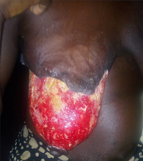
Post debridement.
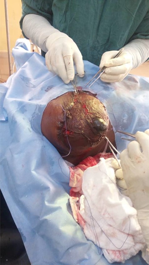
Intraoperative skin graft application.
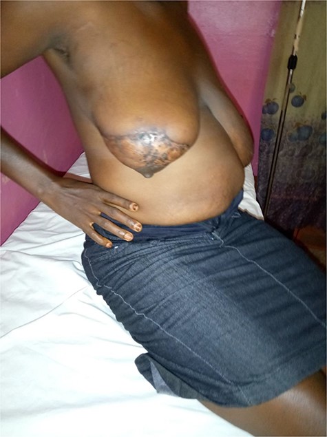
Three months postoperative state.
DISCUSSION
Breast necrotizing fasciitis, although rare, has been a known entity afflicting women either primarily or secondary to known risk factors such as breast engorgement, nipple fissures and previous history of puerperal mastitis [9]. We managed a patient who had breast engorgement following intrauterine fetal death and developed breast engorgement with a background hypoalbuminemia. The patient was resident in a rural area and had poor financial means for general upkeep and feeding. The malnutrition may have predisposed the patient to the infection while breast engorgement may have precipitated it.
In some cases, the cause is not known but is believed to have resulted possibly from trivial fissures or scratches on the lactating breast or otherwise [9,10]. The index case did not breastfeed and so developed engorgement, which was not suppressed prior to breast necrosis. A quick consult to the general surgeons and timely intervention by the general surgeon limited the parenchymal depth of the necrosis, thereby reducing the affectation of the blood vessels coursing through the parenchyma to supply the nipple-areola complex. A similar reason has been adduced for a reported case with nipple sparing [10].
Necrotizing soft tissue infections are often misdiagnosed to the tone of ~41–96%[1,2] They are mistaken for abscess or cellulitis. In the index case it was diagnosed as breast abscess by the gynecological emergency unit but was discovered to be breast gangrene on further review. However, timely intervention helped to limit necrosis.
Excessive milk leakage into the wound was managed with special suspension dressing with multiple vicryl-2 sutures anchored on the edges of the residual breast skin forming a loop for suspending the breast on a drip stand was used. Milk drainage and collection under the graft was further controlled with light pressure dressing. Patient was later put on bromocriptine tablets, which suppressed the milk drainage.
Graft healing was complete in 3 weeks with satisfactory cosmetic improvement. There was good bra symmetry and fair exposure symmetry. The grafted breast improved in shape and projection compared to the more ptotic contralateral breast.
We recommend therefore the commencement of medical suppression of lactation following stillbirths to prevent engorgement and forestall possible necrotizing fasciitis of breast in postpartum period. This with other measures such as improved hygiene, and adequate nutrition, glycemic control as well as immune system support measures may help to prevent this potentially fatal disease.
CONCLUSION
Necrotizing fasciitis of the breast is rare and commonly misdiagnosed resulting in breast loss and even to death. In postpartum period in stillbirths, breast engorgement is a possible precipitating factor and combining with other factors such as malnutrition results to a rapid course of progression with untoward outcome. Early and aggressive surgical intervention measures with medical management of engorgement contribute to improved outcome with nipple areola preservation. Resultant wound is successfully managed with split-thickness skin grafting.
Acknowledgements
We sincerely acknowledge the efforts of the residents in the unit Drs Amaechi Ugbala and Emmanuel Ukpai.
Conflicts of Interest
Authors declare that there was no conflict of interest or competing interest in the report.
Funding
Authors did not receive funds from any source. There was no grant directly or indirectly. All publication expenses would be borne by the authors. There were no study sponsors at any level of the work starting with study design, case analysis/presentation, and manuscript write-up and in the decision to submit for publication.
