


ABSTRACT
Acute calcific periarthritis (ACP) is an unusual cause of monoarticular pain characterised by the deposition of calcium hydroxyapatite in the peri-articular and intra-articular tissues. Although the most commonly affected joint is the shoulder, other joints may be involved, including the wrist. This case report describes a 57-year-old female presenting with wrist pain and swelling associated with amorphous calcification overlying the lunate. The patient improved with the use of non-steroidal anti-inflammatory drugs and splinting. Clinician awareness of the clinical presentation and radiographic features of ACP is important to reduce unnecessary invasive diagnostic procedures such as joint aspiration.
INTRODUCTION
Acute calcific periarthritis (ACP) is a distinct clinical subset of calcium hydroxyapatite crystal deposition disease (HADD). HADD a systemic disease of unknown aetiology characterised by the deposition of calcium hydroxyapatite (HA) in the peri-articular and intra-articular tissues [1]. The most commonly implicated joint is the shoulder, with calcific deposition in rotator cuff tendons representing one of the most common causes of atraumatic shoulder pain [2]. There is also a high rate of asymptomatic calcific deposition, with a reported rate of 7.8% of the general population having radiographic evidence of crystal deposition [3]. Other commonly involved joints include hip, knee, elbow, wrist and hand [4]. Within the wrist, a common site of involvement is the tendon of flexor carpi ulnaris [5].
ACP may initially be misdiagnosed as infection, malignancy or another form of crystal arthropathy such as gout or pseudogout [6]. The clinical course of the condition is variable. Although most cases spontaneously resolve as calcium depositions are absorbed by macrophages, a small minority of patients develop HA crystal arthropathy with synovitis and associated joint destruction. HA crystal arthropathy most commonly affects the shoulder joint, a condition termed the ‘Milwaukee Shoulder’.
CASE REPORT
In this case a 57-year-old female patient with a past medical history of degenerative spondylosis presented to the emergency department with a 1-week history of left wrist pain. There was no history of antecedent trauma. The patient was constitutionally well, with the wrist being globally tender with a limited range of motion. There was palpable firm swelling over the volar aspect of the wrist with no overlying skin changes. Neurovascular examination was normal. Bloods taken on presentation revealed a normal white cell count, serum urate and C-reactive protein. Initial radiographs showed marked, poorly defined soft tissue calcification overlying the palmar aspect of the lunate with no associated bony injury (Fig. 1).
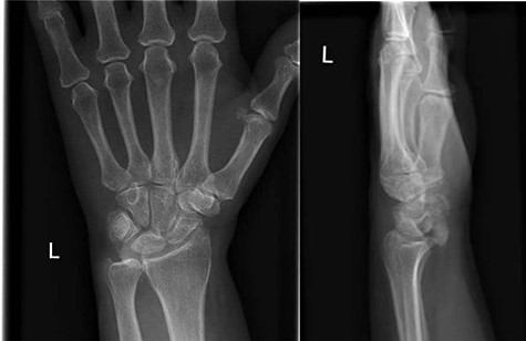
AP and lateral radiographs of the left wrist on initial presentation to the emergency department, showing amorphous calcification overlying the lunate.
The patient was discharged from the emergency department with non-steroidal anti-inflammatories (NSAIDs) and a volar wrist splint. Follow-up review in clinic was scheduled for 2 months later. The patient reported that some swelling and pain persisted however had subsided significantly in the interim. A computed tomography scan was performed which showed almost complete resolution of the amorphous calcification.
DISCUSSION
ACP is an uncommon cause of acute monoarticular pain that is frequently misdiagnosed as septic arthritis or a fracture, particularly when affecting sites other than the shoulder. This case report describes a case of ACP affecting the wrist of a patient, a relatively uncommon site for calcium HA disposition. The calcification had spontaneously resolved within 4 months of initial presentation with splinting and NSAIDs only, without the use of steroids.
Although the clinical presentation of ACP may be similar to a septic arthritis, the presence of amorphous calcification is highly suggestive of the diagnosis. The lack of clinician familiarity with this condition or failure to correctly interpret the radiographic features of ACP may result in unnecessary invasive procedures such as joint aspiration and the use of antibiotics, both of which may result in patient harm. Clinician awareness of the clinical and radiographic presentation of ACP and other forms of HADD is important to reduce the risk of iatrogenic harm whilst investigating what is typically a self-limiting condition.
CONFLICT OF INTEREST STATEMENT
None declared.
FUNDING
None.
