-
PDF
- Split View
-
Views
-
Cite
Cite
Anthony Paolo T Siguan, Marc Denver S Yray, Sebaceous carcinoma of the axilla, Journal of Surgical Case Reports, Volume 2020, Issue 12, December 2020, rjaa513, https://doi.org/10.1093/jscr/rjaa513
Close - Share Icon Share
Abstract
Sebaceous carcinoma is a rare cutaneous malignant neoplasm with an estimated incidence rate of approximately 1–2 per 1 000 000 per year. It rarely presents as an extra-ocular type and it affects patients in between the sixth and eighth decades of life. This is a case of a 31-year-old male with a 10-year history of a right axillary mass with an incisional biopsy of a sebaceous carcinoma. The mass was 10 × 6 cm in largest diameter, erythematous, non-tender and irregularly shaped. The patient underwent wide excision of the right axillary mass with negative margins confirmed by frozen section under general anesthesia. A parascapular fasciocutaneous flap was done in order to provide coverage for the defect. The patient was then discharged improved on the fourth postoperative day. Although there has been no established clinical protocol for the staging, medical and surgical management for extra-ocular sebaceous carcinoma, early diagnosis accompanied with the proper surgical intervention, such as oncologic wide excision with negative margins, were both adequate and paramount to the diagnosis, course and treatment of this patient, given its rarity.
INTRODUCTION
Sebaceous carcinoma is a rare cutaneous malignant neoplasm with an estimated incidence rate of approximately 1–2 per 1 000 000 per year and makes up an estimated 0.05% of all cutaneous malignancies [1]. It is thought to arise from sebaceous glands in the skin, most commonly in the eyelid [2–4]. Thus, these tumors may arise anywhere on the body where these glands exist. This aggressive neoplasm affects patients predominantly between the sixth and eighth decades of life; it is rarely seen in younger individuals, except for those under immunosuppression or radiotherapy [4]. Sebaceous carcinomas have been traditionally classified into two types: the ocular type and the extra-ocular type [5]. Extra-ocular sites for these tumors account for only about a quarter of the cases and usually include the skin of the head and neck regions [6]. This type of cutaneous carcinoma is noted to be very rarely found in the axilla and is often confused with a primary lymphoma or a secondary malignancy from that of the breast [2]. There has been no known published documentation of extra-ocular sebaceous carcinoma in the Philippines.
Significance of the study
Sebaceous carcinoma of the axilla is a rare type of extra-ocular malignancy, and that there has been no report of such incidence in the Philippines. Aside from contributing to medical literature, we can share our experience with this case to the medical community. Being able to discuss the surgical course and management of this case at our local setting will aid surgeons who will encounter this rare type of malignancy.
CASE PRESENTATION
This was a case of a 31-year-old male with a 10-year history of a right axillary mass. The lesion initially began as a 0.5 cm mass on the right axilla noted to be fixed, erythematous, non-tender and notably irregularly shaped. On the interim, condition was just tolerated. Progression of the growing axillary mass prompted consulting at the outpatient department. The irregularly shaped mass was already noted to be 10 cm at its largest diameter. An incisional biopsy was done, which revealed sebaceous carcinoma. The patient was advised for surgery.
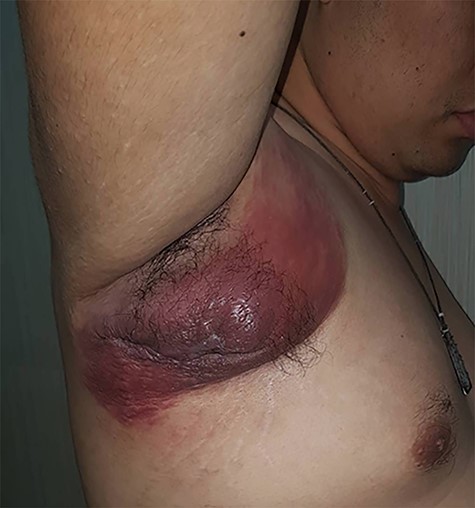
The patient was average built with normal systemic examination findings. On the right axilla, he had a 10 × 6 cm irregularly shaped mass still noted to be erythematous and non-tender (Fig. 1). There were no palpable axillary lymph nodes. Bilateral breasts and the contralateral axilla were unremarkable.

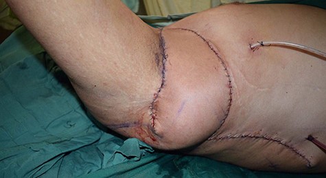
Wide excision of the right axillary mass with frozen section of the margins was done under general anesthesia. Frozen section was done in order to guarantee disease-free margins. This consequently left a full thickness defect around 15 × 30 cm on the right axilla. A parascapular fasciocutaneous flap coverage was subsequently done on the defect (Fig. 2). Patient was then discharged improved on the fourth postoperative day and was advised surveillance follow-up as per outpatient basis.

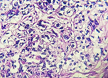
On pathologic gross inspection of the mass, it consisted of skin covered with fibrofatty tissue and was measured to be 19.5 × 10 × 4.5 cm. On sectioning, there was a well-circumscribed pale, tan and rubbery mass, 10 × 6.5 × 6 cm, with areas of hemorrhage and was located 0.3 cm from the nearest basal margins. The microscopic examination of the mass showed sections of malignant neoplasm composed of sheets of highly pleomorphic cells with abundant pale, vacuolated and fluffy appearing cytoplasm with distinct boarders and small to large vesicular nuclei with prominent nucleoli, while some nuclei appeared pyknotic (Fig. 3). The surrounding stroma was fibrous, with heavy mononuclear infiltration. The neoplasm showed infiltrative boarders. Sections of the skin were free of tumor. No epidermal tissue was seen in continuity with the tumor. There was no clefting between the stroma and the neoplastic cells. The final biopsy of the mass revealed poorly differentiated sebaceous carcinoma with all surgical margins negative for tumor; furthermore, two axillary lymph nodes were negative for tumor metastasis.

Section of the mass on hematoxylin and eosin stain at high power field magnification.
CASE DISCUSSION
Sebaceous carcinoma is a rare, aggressive neoplasm that most commonly presents as an ocular or peri-ocular mass [10]. Among the extra-ocular types of this malignancy, the parotid gland is the most common predilection; however, the patient presented with this extra-ocular type of neoplasm at an atypical location which was at the axilla. Furthermore, this neoplasm rarely presents in the younger population unless with a history of immunosuppression or exposure to radiation [4]. This patient is only 31 years old. There has been a relative increase of incidence with this type of malignancy among the Asian population, which is concurrent since the patient is of Filipino descent. The most important prognostic factor for sebaceous carcinoma is early diagnosis. The mortality rate rises from 14 to 38%, if the diagnosis is made after the first 6 months [7]. Surgical therapy is considered the primary treatment for this tumor [8–10]. The patient underwent wide excision of the right axillary mass with a parascapular fasciocutaneous flap under general anesthesia. Histopathologic examination is key for a definitive diagnosis. Early diagnosis and surgical intervention are crucial to improve survival, given its highly metastatic potential [8–10].
To date, there has been no established clinical protocol or guidelines for both staging and management for extra-ocular sebaceous carcinoma. No recommended surveillance measures have also been established to screen for associated syndromes of this type of malignancy.
CONCLUSION
Although there has been no established clinical protocol for the staging, medical and surgical management for extra-ocular sebaceous carcinoma, early diagnosis accompanied with the proper surgical intervention, such as oncologic wide excision with negative margins, were both adequate and paramount to the diagnosis, course and treatment of this patient given its rarity.


