


Abstract
Caudal cutaneous appendage is a rare condition. According to association with underlying spinal dysraphism, it can be classified into true or pseudotails. Management and prognosis depends closely on spinal anomaly. Fewer than 40 cases of true tail were reported. We describe a rare case of true tail in a newborn explored and operated in our unity.
INTRODUCTION
Caudal cutaneous appendage is a rare condition that aroused interest since 1880s [1]. It is defined by midline protrusion at the lumbosacrococcygeal region. It can be classified on true human tail or a pseudotail if associated to underlying spinal dysraphism, which has a different prognosis and requires specialized management [2, 3].
True human tails are very rare, with fewer than 40 cases reported to date [4].
We report a healthy 10-day-old male newborn who was referred to our surgical unity with a cutaneous appendage araising from the lumbosacral discovered on post natal.
CASE REPORT
A 10-day-old male neonate was referred to our paediatric surgical department in the month of September 2019 with a cutaneous appendage arising from the lumbosacral region which had been discovered at birth. The neonate had been born by vaginal delivery and the mother had no history of illness, radiation exposure or drug intake during the antenatal period. In addition, there was no history of congenital anomalies in any of the family members or siblings.
Clinical examination of the new born found a 3.4 kg baby, with no dysmorphia, no anomaly of the limbs or genital organs. The lumbosacral examination found a 6-cm long-shaped soft tissue appendage arising and hanging down from the mid-sacral region, which was completely covered by skin and had no spontaneous movement (Fig. 1).
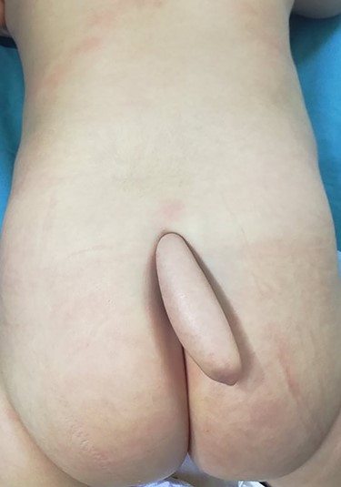
Clinic aspect of human tail.
The mass of the appendage was f firm and was not sensitive. No bone or cartilage could be felt (Fig. 1). No abdominal mass or anal anomaly was detected.
A sacrococcygeal teratoma was suspected an alphafoetoprotein dosage was performed and it was normal.
Magnetic resonance imaging (MRI) of the spine showed no evidence of spinal dysraphism, suspecting the diagnosis of a true human tail (Fig. 2).
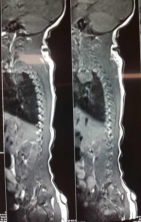
MRI showing connection with soft tissue.
Surgical management was performed: a midline lozenge incision was done encircling the base of appendage. The subcutaneous lipoma was excised and there were no connection with soft tissue or sacroma as shown in the MRI (Fig. 2).
Microscopic examination confirmed the absence of any bony or cartilaginous structures. And showed lipoma without signs of malignancy or nerve tissue. The postoperative period was uneventful.
DISCUSSION
First described as a mythical Greek Mythology by both Virchow (1884) and Miller (1881) or even legends, anecdotes and tales blended with superstition (Gould and Pyle, 1897), several scientific and embryological theories have been proposed since that time [1, 2].
The most retained theory is that human tail results from incomplete regression of the most distal end of the normal embryonic tail found in the developing human foetus [4]. In 1998, Lu et al. [5] suggest that the presence of a human tail is an abnormality in embryonic development, rather than a regression in the evolutionary process.
According to Greco TL et al., it is possible that mutations resulting in increased up regulation of the WNT3A gene may result in retention of the embryonic tail in humans. However, further genetic studies are necessary [6].
True human tails are not inherited, but familial cases have been described and in one case, three female generations in the same family were born with true human tails [7]. In our patient, there is no similar case in the family.
Human tails may be associated with other congenital anomalies in 29% of cases and review of the literature showed spina bifida to be the most frequent coexisting anomaly with both true tail and pseudotail [8]. In our case, the MRI invalidated any associated spine anomaly.
According to Dao and Netsky, there are features to distinguish true tails from pseudo tails. A true human tail is a distal skin-covered boneless midline protrusion which is a remnant of the embryonic tail, containing striated muscle, adipose and connective tissue with blood vessels, nerve fibres, ganglion cells and mechanoreceptors [2, 4]. Pseudo tails is an anomalous prolongation of the coccygeal vertebra, lipoma, teratoma or condrodystrophy [9] and it is often associated with the spinal and coccygeal anomalies. Our patient presented with true tail without spinal anomaly.
The distinction between the true tail and pseudotail on clinical examination is delicate and despite a normal neurological examination, and a normal x-ray of spine, a spinal MRI should be performed preoperatively to classify the tail. Surgical management and prognosis can change in both situations.
CONCLUSIONS
The caudal appendage or ‘human tail’ is a rare anomaly. Its recommended to be watchful when dealing with some of those lesions because its innocent-looking appearance may induce the surgeon to perform a simple excision of the malformation. However, special care should be taken to exclude any serious associated anomaly.
Prognosis is excellent in case of true tail, for pseudo tails, neurological deficiency may occur.
CONFLICT OF INTEREST STATEMENT
None declared.
FUNDING
None.
