


Abstract
Caecal volvulus represents 25–40% of all colonic volvulus. Symptoms include abdominal distension, constipation, nausea and vomiting where it may be intermittent. Abdominal X-rays and computed tomography (CTs) may help with diagnosis and recommended treatment is resection of mobile caecum. A 70-year old comorbid woman with previous open bariatric surgery and known incisional hernia presented with symptoms of bowel obstruction. CT showed caecal volvulus contained within the ventral hernia confirmed intra-operatively. Patient recovered well and was discharged on Day 6 of admission. This is the second case described in literature of a caecal volvulus occurring in an incisional hernia. The altered normal anatomy may have contributed to caecal mobility. Diagnosis of caecal volvulus can be challenging, more so in the presence of a more clinically apparent pathology. We present a second known case of caecal volvulus in a giant incisional hernia, where there were unique challenges to management.
INTRODUCTION
We present a unique case of caecal volvulus that occurred within this patient’s large ventral hernia; this has been only reported once before in the literature [1] is estimated that caecal volvulus accounts for 25–40% of all colonic volvulus [2]. The overall mortality is ~6.7%, with the presence of peritonitis, bowel gangrene and necrosis significantly increasing the mortality rate [3]. Despite the low rate of colonic volvulus causing bowel obstruction (1.9% of all bowel obstructions), the incidence of caecal volvulus is increasing by ~5.5% a year [3].
Most patients present with symptoms of abdominal distension, cramping pain, constipation, nausea and vomiting [2, 4, 5]. Up to 50% may present with intermittent symptoms where caecum spontaneously torts and detorts [6, 7] prior to full torsion. This is a non-specific finding and makes diagnosis difficult. Laboratory findings are generally non-specific but point towards metabolic compromise due to the sequelae of bowel obstruction or vascular compromise [4]. Abdominal X-rays may show caecal dilation, an absence of gas in distal colon and small bowel dilatation. Abdominopelvic computed tomography (CT) scans are the imaging modality of choice as it approaches 100% sensitivity and >90% specificity [4], classic findings would include the whirl sign, as well as a coffee bean sign where the caecum is displaced to the left upper quadrant [2, 8, 9].
Various methods of managing caecal volvulus are in the literature. Colonoscopic reduction has been described, but the efficacy is ~30%. Combined with the risk of perforation and potential delays in the event of unsuccessful reduction, it is not recommended as initial treatment [2, 5]. A variety of operative approaches have also been described, including caecopexy, caecostomy or resection. Resection of the mobile caecum is the recommended treatment of choice [2, 4, 5], as caecopexy and caecostomy are associated with increased morbidity and mortality [4] as well as high recurrence rates [2, 3]. It is accepted that bowel continuity can be restored in a single operation but degree of illness and patient factors may necessitate a stoma [3, 5].
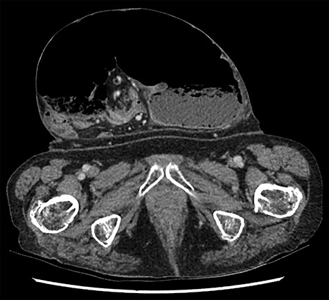
Caecal volvulus, showing distended caecum within ventral hernia showing mesenteric swirling (axial).
CASE REPORT
A 70-year-old woman presented with colicky abdominal pain and increasing size of a large ventral hernia associated with 3 days of obstipation and vomiting. Her past history includes chronic obstructive pulmonary disease (COPD), previously requiring home oxygen but ceased due to non-compliance, lower limb venous thromboembolism (VTE), chronic back pain and polymyalgia requiring long term opiates and steroids. She had a large incisional hernia for at least 7 years following open gastric bypass surgery 40 years prior for weight loss, but due to her co-morbidities and the relative wide neck of the hernia where strangulation of the contents were deemed unlikely, she had previously been deemed not suitable for elective repair.
Her vital signs on presentation were respiratory rate of 20, SpO2 of 94%, BP 142/87, pulse 93 and temperature of 36.7. The abdominal examination revealed a large incisional hernia, ~20 cm in diameter with a 10 cm neck. This was tender without peritonism. She had an elevated C-reactive protein (CRP) but blood results were otherwise unremarkable.
A clinical diagnosis of bowel obstruction was made; she underwent a CT abdomen/pelvis with contrast, which demonstrated a large bowel obstruction within the hernia not caused by the hernial defect but thought likely due to volvulus of the caecum (Figs. 1 and 2).
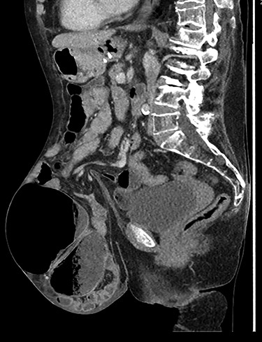
Caecal volvulus through wide-necked abdominal wall defect with small amount of free fluid in pelvis (sagittal).
After discussion with the patient regarding her high risk of morbidity and mortality, decision was made to operate. She underwent a midline laparotomy and the hernia sac was entered, which revealed a caecal volvulus with complete 360° rotation on the vascular pedicle but with viable bowel. A limited ileocolic resection was performed with formation of an ileostomy. Tension-free primary closure of the abdominal wall was successful. She remained intubated overnight in ICU, was extubated the next day, and otherwise recovered uneventfully. She was discharged Day 6 post admission.
DISCUSSION
Risk factors for developing caecal volvulus for the patient were being female and having had previous abdominal surgery. It is possible that due to the long-standing herniation of the colon from the abdominal cavity, resulted in elongation of the mesentery combined and increased caecal mobility due to losing fixation points causing her to be at increased risk developing caecal volvulus, a view shared with the authors of the first case report [1]. Similarities to the previously reported case that are noted include the history of prior surgery and presentation of patient to the emergency department >24 h after symptom onset. However, it is fortunate that our patient has remained clinically well throughout her course of treatment with bowel viability maintained, unlike the previous case, and with that, a single stage operation was able to be performed. The operative approach in this case differs to a standard repair of a large incarcerated incisional hernia as resection of mobile caecum was required to prevent recurrence of volvulus.
There have been various challenges to the management of the patient. The first of which is the challenge of making the right diagnosis. Caecal volvulus often presents non-specifically which impairs the ability to make a clinical diagnosis [4, 9]. Furthermore, the presence of a more prominent clinically apparent pathology of the large incisional hernia in this case made diagnosis more challenging.
A common severe complication of large hernia surgery is respiratory failure due to shifts in intra-abdominal and intra-thoracic volumes [10]. This has been considered a major risk with her pulmonary disease and in addition, her chronic immunosuppressed state has precluded her for an elective repair. It is fortunate that this patient has progressed through her immediate post-operative course well without the need for prolonged respiratory support and this has challenged our initial thoughts of her being of too high of a risk for elective surgery, which may have prevented the development of a mobile caecum and hence, this acute admission. It is to note that the patient’s abstinence from smoking for the last 3 months may have helped her recovery and decisions are easily made retrospectively.
As a conclusion, this highlights the importance of balancing the risk versus benefits of hernia repair and the presence of caecum or sigmoid colon in large ventral hernias may necessitate repair due to risk of volvulus.
CONSENT
Written informed consent was obtained from the patient for the publication of this case report and accompanying images. A copy of the written consent is available for review by the Editor-in-Chief of this journal upon request.
CONFLICT OF INTEREST
None declared.
FUNDING
None.
AUTHOR CONTRIBUTION
Y.T.: Admitting patient from emergency department, literature review, and writing of paper; A.F.: performed patient surgery and peri-operative care, selection of images, and editing and formatting paper for publication; M.T.: primary admitting consultant, performed surgery for patient, review and formatting of paper for publication.
