


Abstract
Nasogastric tubes (NGTs) have long been used for various indications, most commonly to decompress the stomach of its contents in intestinal obstruction or after abdominal surgery, to provide enteral feeding or to allow enteral liquid medication administration. Recently greater importance has been given to the correct placement NGTs to avoid serious complications. We present a case of a spontaneously knotted NGT that was identified and safely removed without complications, but which may have resulted from suboptimal placement. We discuss this case to raise awareness of this complication and how to minimize the likelihood of it happening and improve patient outcome.
INTRODUCTION
Nasogastric tubes (NGTs) are widely used in the medical practice. As with any other invasive procedure, they are not without complications.
Complications associated with NGT are usually either during NGT insertion or removal.
Spontaneous true knot formation in the tube is rarely encountered but if undiagnosed can cause unanticipated trauma.
Factors contributing to NGT knotting are related to insertion as well as removal.
CASE REPORT
A 58-year-old female presented to the Accident and Emergency department with a 1-week history of nausea, vomiting, constipation, abdominal pain and distension.
General examination was unremarkable and vital signs were normal. Abdominal examination revealed distension with rebound tenderness in the left iliac fossa and suprapubic region. Digital rectal examination revealed hard stool in the rectum.
Initial laboratory investigations revealed leucocytosis (white blood cells, 18.4 × 10⁹/L), sodium 137 mmol/L and potassium 3.6 mmol/L, hemoglobin 12.5 g/dL, platelets 312 × 109/L, prothrombin time 17.1 s, activated partial thromboplastin time 28.3 s, amylase 17 U/L, total bilirubin 20 mg/dL, alanine aminotransferase 16 U/L, alkaline phosphatase 82 U/L, total calcium 2.37 mmol/L and albumin 39 g/L.
Abdominal X-ray revealed impacted stools with possible obstruction. Computed tomography scan suggested an inflammatory process likely due to diverticulitis of the distal sigmoid with localized perforation. Emergency laparotomy was carried out after fluid resuscitation confirming stercoral perforation for which a Hartman’s procedure was performed.
Before the operation, an NGT was inserted through the nostril after application of lubricant gel to decompress the stomach, fixed to the nose at 95 cm. (Fig. 1-2).
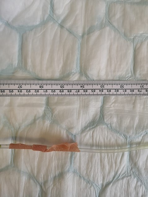
Photo showing NGT proximal end taped to the nose at 95 cm.
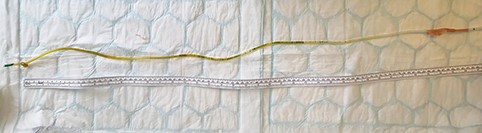
Photo showing whole NGT.
Postoperatively, the NGT continued to function for a couple of days. The patient had a smooth post-operative recovery and on the fourth postoperative day was able to tolerate fluids such that the surgical team advised to remove the NGT. The nurse looking after the patient tried to pull the NGT out but the tube end stuck at the nose. Even after gentle manipulation by different surgical team members, the NGT failed to be removed.
She was referred to ear, nose, and throat (ENT) specialist who managed to remove the tube using a local anesthetic spray to oropharynx and gentle bedside manipulation. The tube was found to have a knot 4 cm from its distal end. (Fig. 3).
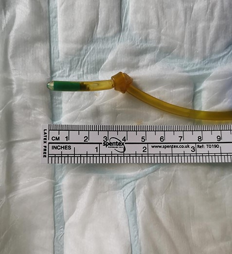
Photo showing the knot 4 cm from NGT distal end.
The patient was discharged the following day with no complications.
DISCUSSION
NGTs have been used for a long time for various indications, most commonly to decompress the stomach of its contents, usually after bowel obstruction, abdominal surgery or mechanical ventilation, to aspirate ingested toxic materials, to provide enteral feeding or for liquid medication administration.
Iatrogenic complications in medicine are increasing, in line with a general increase in invasive procedures. NGT insertion and removal is a routine, straight forward invasive procedure, most of the time carried out by trained registered nurses Awareness of possible complications related to the NGT will help improve patient outcomes.
NGT-related complication scan is divided into nasal complications (including ulceration, epistaxis), esophageal complications (including erosion and perforation), pulmonary complications (including aspiration, bronchial and lung injury) and tube-related complications (including tube breakdown and knotting).
NGT knotting is a rare and often overlooked complication with the potential to cause significant trauma on tube removal if unrecognized [1].
Although knotting of the NGT has been reported in the literature, its incidence appears to be very low [2].
Risk factors include the length of the tube, smaller diameter tubes, insertion deep into the stomach and interference with an endotracheal tube in the intubated patient [3–4].
It is reported that knotting of the tube occurs during insertion and that the traction during retrieval tightens the knot. Our case supports this theory because the tube continued to function while it was in situ. The resistance experienced during the removal of the tube increases progressively as the catheter passes through the narrowest part of the esophagus. [5–6]
Egan et al. [7] reported a similar case where upon attempting to remove the tube, the patient experienced severe pain and developed epistaxis. In their example, the ENT surgeon was able to advance the NGT, cut the external distal portion and retrieve the remainder through the oropharynx.
NGT-related complications are usually preventable. Poor reporting of these complications may slow adoption of effective preventative guidelines.
This case report serves as a reminder to clinicians of a rare but possible complication which is in part preventable.
Large NGT caliber, as well as appropriate NGT length (based on external measurement from the tip of the nose to a point halfway between the xiphoid and the umbilicus), can decrease the chances of NGT knotting.
AUTHOR’S CONTRIBUTIONS
All authors contributed to patient’s management, literature review as well as manuscript editing.
CONFLICT OF INTEREST STATEMENT
None declared.
PATIENT’S CONSENT
Written informed consent was obtained from the patient to include images in the article.
