-
PDF
- Split View
-
Views
-
Cite
Cite
Apinan Rongviriyapanich, Adenocarcinoma of jejunum, Journal of Surgical Case Reports, Volume 2018, Issue 8, August 2018, rjy234, https://doi.org/10.1093/jscr/rjy234
Close - Share Icon Share
Abstract
Although gastrointestinal malignancy is still the major concern of health problems in Worldwide and Thailand, but small intestinal malignancy is extremely rare. The location of small intestinal malignancy is duodenum (73.6%), jejunum (13.2%) and ileum (13.2%). The diagnosing of small intestinal malignancy usually delays due to inaccessible of esophagogastroduodenoscopy especially jejunum and ileum causing poor prognostic outcomes. We reported our case of jejunal adenocarcinoma.
INTRODUCTION
Although gastrointestinal malignancy is still the major concern of health problems in Worldwide and Thailand, the small intestinal malignancy is extremely rare. Only 0.6% of all malignant patients in the USA are small intestinal malignancy [1]. The National Cancer Institute of Thailand reported eight new cases of small intestinal malignant patient in Thailand around 2015 and only three cases had histopathological examination showing adenocarcinoma [2].
The location of small intestinal malignancy is duodenum (73.6%), jejunum (13.2%) and ileum (13.2%) [3].
The diagnosing of small intestinal malignancy usually delays due to inaccessible of esophagogastroduodenoscopy especially jejunum and ileum causing poor prognostic outcomes. The 40–60% of all patients can be cured and only 32% are non-metastatic lesions [3]. The 5-year survival rates of small intestinal malignant patient is 67.5% [1].
We reported our case of jejunal adenocarcinoma.
CASE REPORT
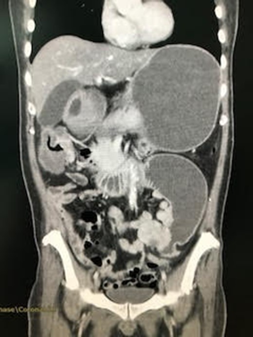
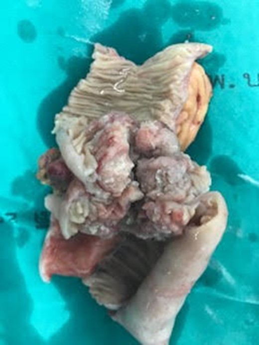
A Thai male patient, aged 46 years, presenting with chronic abdominal pain for 3 months. He frequently visited hospital in 3 months about chronic abdominal pain with unidentified cause—recurrent hyponatremia. He underwent upper gastrointestinal endoscopy and ultrasonography of upper abdomen but showed within normal limit of both examinations, so he was treated as chronic dyspepsia and symptomatic treated about hyponatremia but he still not improves. In admission day, he re-visited hospital with abdominal pain and his blood chemistry showed hyponatremic hypokalemic metabolic alkalosis, so gastric outlet obstruction was provisional diagnosis. Saline loading test showed delayed gastric emptying time. Patient was sent for computed tomography of abdomen because previous upper gastrointestinal endoscopy showed normal study. Computed tomography resulted in 5 cm long, enhanced wall thickening at duodenojejunal junction in left lower abdominal region, abutting adjacent sigmoid colon with preserved fat plane separation and markedly dilatation of the proximal duodenum and stomach is detected as in Fig. 1. The diagnosis of obstructed proximal jejunum was made and exploratory laparotomy was decided. In operative field, a 5 cm Cauliflower mass at proximal jejunum, 15 cm distal to ligament of Treitz, causing proximal duodenal dilatation and distal small intestines collapse was found as in Fig. 2. No liver, omental or peritoneal nodule was found. Radical segmental proximal jejunal resection (with 5 cm proximal and distal margins, with mesentery that vascular supplied resected jejunal segment) with primary small intestinal anastomosis was done. Operative time was 48 min and estimated blood loss was 50 mL. After operation, patient fully recovered but superficial surgical site infection was found and pus culture showed Proteus mirabilis and Escherichia coli. Pathologic examination showed well differentiated adenocarcinoma of jejunum with extension into perienteric fatty tissue, free all resected margins, and reactive hyperplasia of three lymph nodes. The diagnosis of Adenocarcinoma of the jejunum (pT3N0M0—stage II) was made and patient was referred to the oncologist for possibility of adjuvant therapy but the oncologist decided not to give patient adjuvant therapy. Patient remains healthy after 9 months of operation.

CT abdomen showed jejunal mass and proximal stomach and small intestine dilatation.

DISCUSSION
Although gastrointestinal malignancy is still the major concern of health problems in Worldwide and Thailand, the small intestinal malignancy is extremely rare. Only 0.6% of all malignant patients in the United States are small intestinal malignancy [1]. The National Cancer Institute of Thailand reported eight new cases of small intestinal malignant patient in Thailand around 2015 and only three cases had histopathological examination showing Adenocarcinoma [2].
The location of small intestinal malignancy is duodenum (48–73.6%), jejunum (13.2–31%) and ileum (13.2–21%) [3, 4]. From the statistics, jejunal malignancy is extremely rare.
The clinical presentations of patients with primary small intestinal adenocarcinoma were abdominal pain (71.7%), abdominal distension (17.0%), bleeding (5.7%) and jaundice (13.2%) from the study of Hong et al. [3]. In contrast, Duerr et al. [4] showed that clinical presentation of their patients were abdominal discomfort (33%), obstruction (27%), gastrointestinal bleeding (26%), jaundice (2%) and asymptomatic (12%).
The diagnosing of small intestinal malignancy usually delays due to inaccessible of esophagogastroduodenoscopy especially jejunum and ileum. Distal located small intestinal adenocarcinoma was diagnosed with laparotomy (85.7%) and through computerized tomography (14.3%) [3].
Our case presented in the same presentation and the same way of diagnostic modality as reported by Prabhu et al. [5].
The difficulty of diagnosis of small intestinal adenocarcinoma caused poor prognostic outcomes. Hong et al. [3] presented that most of patients were diagnosed when tumor was higher stage (28.3% in stage 3 and 41.5% in stage 4) consistent to study of Verma and Stroehlein [6] that showed distant metastasis in 43% of patients. The 40–60% of all patients can be cured and only 32% are non-metastatic lesions. The 5-year survival rates of small intestinal malignant patient from SEER database is 67.5% [1]. Unfortunately, Hong et al. [3] showed the poorer outcomes in 3-year overall survival rates as 44.1%.
Surgical resection is the only curative option for small intestinal adenocarcinoma. Despite of this, the surgical procedure for jejunal adenocarcinoma remains controversial. Fronticelli suggest a segmental resection on the left side of the mesenteric vessels to protect the blood supply of the duodenal stump and identified radical resection of this disease as having a proximal resection margin of more than 2 cm long and extended lymphadenectomy is recommended [7].
CONCLUSION
The jejunal adenocarcinoma is extremely rare. The diagnosing of this usually delays due to inaccessible of esophagogastroduodenoscopy causing poor prognostic outcomes. Surgical resection is the only curative option.
CONFLICT OF INTEREST STATEMENT
None declared.


