-
PDF
- Split View
-
Views
-
Cite
Cite
Zishan Jetha, Michael Lisi, Prolapsed fundic gastric polyp causing gastroduodenal intussusception and acute pancreatitis, Journal of Surgical Case Reports, Volume 2018, Issue 7, July 2018, rjy139, https://doi.org/10.1093/jscr/rjy139
Close - Share Icon Share
Abstract
An 86-year-old female presented with a 6-month history of recurrent intermittent epigastric abdominal pain, postprandial fullness with nausea, vomiting, anemia and a 15-pound weight loss. A large fundic gastric polyp was intussuscepting into the duodenum causing intermittent compression and obstruction of the ampulla of Vater leading to acute pancreatitis. An overview of the clinical presentation, diagnosis and management of this entity, in addition to a review of the literature is provided.
INTRODUCTION
Gastric polyps are rare lesions. They are usually found incidentally during upper gastrointestinal endoscopies. Although mostly asymptomatic, larger lesions have the potential to prolapse through the pylorus, creating a setting for gastric outlet obstruction and acute pancreatitis. Not only are these lesions a recognized source of morbidity, but larger lesions are frequently associated with a component of high-grade dysplasia with the potential for malignant transformation. As a result, consideration needs to be made for further investigation and resection if necessary.
CASE REPORT
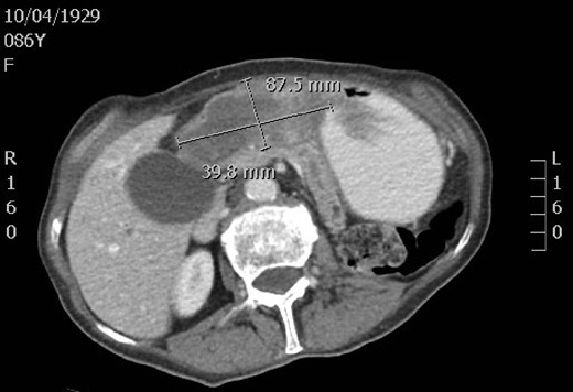
An 86-year-old female initially presented to the emergency department with a 6-month history of recurrent intermittent epigastric abdominal pain, postprandial fullness with nausea, vomiting, anemia and a 15-pound weight loss. Her past medical history was significant for hypertension and clear cell renal cell carcinoma. A contrast-enhanced computed tomography scan revealed a large, irregular, solid mass in the gastric fundus measuring 8.8 × 4.0 by 3.7 cm. Mild pancreatic duct dilation was also appreciated. During an initial gastroscopy, great difficulty was encountered while trying to pass through the pylorus into the duodenum. A barium swallow study revealed gastric fundal thickening and there was markedly delayed emptying and gastric outlet obstruction noted. The patient’s symptoms improved with conservative management of fluids and analgesics but her symptoms relapsed when attempts were made to advance her diet.
The patient returned to the emergency department ~1 month later with non-remitting epigastric pain and vomiting. There was no history of previous pancreatitis, alcohol abuse, metabolic disease, or previous trauma. She was, however, known to have cholelithiasis. On clinical examination, she was afebrile and was tender in the epigastrium. She had a negative Murphy’s sign. Laboratory assessments revealed a hemoglobin of 108 g/L, amylase of 357 U/L and lipase of 8700 U/L. Otherwise, her white cell count, electrolytes and liver function tests were unremarkable. A repeat contrast-enhanced computed tomography scan revealed interval dilation of the main pancreatic duct and gastroduodenal intussusception with a lead point suspected to be associated with the fundic mass. A repeat gastroscopy revealed a large polypoid mass originating from the fundus of the stomach and protruding past the pylorus into the duodenum. It was difficult to maneuver into the duodenum once again and the mass could not be reduced endoscopically.
The patient was reviewed at multidisciplinary tumor boards and it was felt that the mass was not endoscopically resectable. As a result, the patient underwent a mini laparotomy and a gastrostomy was performed. The polypoid mass was resected using a gastrointestinal anastomosis stapler and the staple line was then oversewn. The patient tolerated the procedure without any complications and was discharged from hospital a few days later tolerating a full diet. The final pathology revealed pyloric gland adenoma with positive foci of high-grade dysplasia, and negative for invasive carcinoma (Fig. 1).

A computed tomography scan revealing a large, irregular, solid mass in the gastric fundus.
DISCUSSION
Gastric polyps are lesions that project above the mucosal plane into the lumen of the stomach. They are usually uncommon and identified incidentally during 3–5% upper gastrointestinal endoscopies [1]. Fundic gland polyps (80%) and gastric hyperplastic polyps (19%) comprise the majority of all benign gastric polyps, while adenomas and neuroendocrine tumors both comprise <1% of all gastric polyps [2]. Most patients are asymptomatic but infrequently, complications such as bleeding from ulceration or gastric outlet obstruction may occur. The reported patient had a large broad-based polypoid mass projecting from the gastric fundus through the pylorus and into the duodenum causing obstruction of the ampulla of Vater. This lead to clinical and radiographic evidence of acute pancreatitis. Her symptoms were relapsing in nature, presumably due to intermittent compression against the hepatopancreatic outlet.
There have been only a few cases of gastric polyps prolapsing through the pylorus causing gastric outlet obstruction and acute pancreatitis. Even fewer report an adenomatous polyp originating from the fundus, as in the present case [3, 4]. Most adenomas are solitary, exophytic lesions which can be sessile or pedunculated and usually measure <2 cm. Larger adenomas are more frequently associated with a component of high-grade dysplasia and a significant proportion contain foci of malignant transformation.
A minority of gastric adenomas show morphologic characteristics of gastric foveolar or pyloric gland-type epithelium as in the present case. Pyloric gland adenomas tend to occur more frequently in females with an average age of 75 years [5]. In the stomach, the gastric body is the most common location, followed by the gastric transition zone, antrum and cardia. Grossly, they appear polypoid, dome-shaped or as fungating masses. They are associated with autoimmune metaplastic atrophic gastritis, Helicobacter pylori gastritis, chemical gastritis and are on average 1.6 cm at the time of diagnosis [6]. Since they are pre-invasive neoplasms with potential for progression to adenocarcinoma, gastric adenomas must be treated by local excision. In addition, since they may be accompanied by coexistent carcinoma elsewhere in the stomach, a thorough evaluation of the remaining stomach and follow-up screening may be necessary.
Intussusception in adults is a relatively rare condition and accounts for only 1–3% of patients with intestinal obstruction [7]. Gastric intussusceptions are even less prevalent. About 90% of the intussusceptions in adults occur in the small or large bowel, while the remaining 10% involve the stomach or surgically made stomas [8]. They tend to occur secondary to mobile pedunculated lesions, which act as lead points for anterograde prolapse of gastric mucosa. They are usually associated with benign and malignant lesions including smooth muscle tumors, hyperplastic and adenomatous polyps, lipomas, and rarely malignant masses [9]. The symptoms of gastroduodenal intussusception usually are non-specific and may consist of abdominal pain, dyspepsia, nausea, vomiting and vary directly with the ability to reduce the intussusception. Acute pancreatitis can be triggered by a temporary or permanent blockage of the pancreatic duct. Our patient’s symptoms were intermittent in nature and partially resolved on both occasions post endoscopic intervention, despite the obstructing mass not being completely reduced.
Currently, there are no formal guidelines in the literature for the management of large gastric polyps. In a study reviewing 39 cases of gastric polyps leading to gastric outlet obstruction, the median size of polyps removed endoscopically was 3 cm, while the median size of surgically removed polyps was 6 cm [10]. The largest polyp removed endoscopically was 8 cm. However, initially two-thirds of the polyp was snared and then the remainder was excised during a subsequent intervention. In the case presented, consideration was made to initially excise the fundic polyp endoscopically but it was felt to be too large. Laparoscopic gastrostomy for polyp excision was also considered but ultimately, a small laparotomy was favored.
CONCLUSION
Although gastric polyps are relatively rare lesions, large gastric lesions have the potential to prolapse through the pylorus, creating a setting for gastric outlet obstruction and acute pancreatitis. The case presented here is unusual in that the broad-based polypoid mass was found to be a pyloric gland adenoma originating in the fundus leading to gastroduodenal intussusception. Because these lesions are frequently associated with high potential for malignant transformation, they require a judicious clinical approach. Although smaller lesions may be amenable to endoscopic resection, a surgical intervention is likely more suitable for larger lesions. Further investigations are needed to help clarify the boundaries of endoscopic versus surgical resection.
CONFLICT OF INTEREST STATEMENT
None declared.


