-
PDF
- Split View
-
Views
-
Cite
Cite
Eleftheria Mavrigiannaki, Ioannis Fesatidis, Evgenia Kalogridaki, Ioannis-Petros Katralis, Dimitrios Filippou, Panagiotis Skandalakis, Dionisios Vrochides, Synchronous nephrectomy and cavoatrial tumor thrombectomy under normothermic extracorporeal circulation and beating heart, Journal of Surgical Case Reports, Volume 2018, Issue 5, May 2018, rjy095, https://doi.org/10.1093/jscr/rjy095
Close - Share Icon Share
Abstract
Formation of tumor thrombus is an occasional manifestation of renal cell carcinoma (RCC). Intravascular invasion of the renal vein and thereafter the inferior vena cava (IVC) might in very rare cases extend into the cardiac chambers. The subtle course and symptoms of such cases alongside with the engagement of vital anatomical structures marks them as a diagnostic and therapeutic challenge. Aggressive surgical intervention has proven to be critical for survival rates in such cases; however total synchronous resection remains a challenge for the surgical team and a debate for the medical community. Following we report the case of a 66-year-old male who was diagnosed with a RCC of the right kidney accompanied by a tumor thrombus extending into the right atrium, after he suffered a presyncope episode. The patient underwent a radical en bloc nephrectomy and tumor thrombectomy under extracorporeal circulation with beating heart.
INTRODUCTION
Renal cell carcinoma (RCC) is characterized by its high metastatic index and its propensity to invade intravascular and generate tumor thrombi. Approximately 15% of all RCCs will invade the inferior vena cava (IVC) but only 1% of them will extend supradiaphragmatic into the right atrium, classified as a Level IV tumor thrombus according to the Neves and Zincke system [1, 2]. Total surgical resection is the gold standard of therapy in these patients [3]. Average survival without any kind of surgical intervention is 5 months [4]. Surgical removal launches 5-year survival from 40 to 60% [4]. Due to the rarity of such patients there are still many controversies regarding the most appropriate surgical technique. The aim is to limit complication and mortality rates thus improving prognosis. Precise tumor staging studies, collaboration of an experienced multidisciplinary team and patient’s consent are prerequisites for therapeutic planning.
CASE REPORT
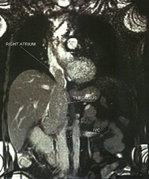
A 66-year-old male, former smoker with a history of hypertension, chronic obstructive pulmonary disease and glaucoma was delivered to the emergency department after a presyncope episode. The patient mentioned episodes of diarrheic melaenas over the last 2 weeks, a progressively worsening dysthymia over the last 2 months and a constant pain of the right lower lumbar region of more than five months that was diagnosed as a hernia. ECG showed sinus rhythm with frequent atrial ectopics. The clinical examination was without special findings and malaena was not clinically confirmed. Vital signs were measured within normal limits. Laboratories revealed anemia (Hct 30.4%, Hb 9.8%), mild elevation of liver enzymes (gGT 197, ALP 186) and CRP (14.1 mg%). Cardiac markers and fecal occult blood test were negative. An abdominal ultrasound revealed a heterogenous mass (6.8 × 6.7 cm2) on the upper pole of the right kidney and a tumor thrombus extending to the IVC. The CT scan of the abdomen and the thorax confirmed the diagnosis of renal mass with cavoatrial tumor thrombus. Pre-surgical staging with MRI and angiography revealed no other sites of pathology or metastasis (Fig. 1).

The patient underwent a radical en bloc nephrectomy and thrombectomy under extracorporeal circulation in normothermia and beating heart. The patient remained on ICU for 7 days and on the fourth day, following oedema of the right lower leg, a femoral and iliac vein thrombus was discovered. This was corrected surgically and no other complications were incurred. He was discharged on Day 19. Two years postsurgery a possible retroperitoneal tumor was detected and removed by a median laparotomy. Histology did not reveal any features of malignancy. The patient, 4 years after initial surgery, is under oncological follow-up, receives targeted therapy and no other sites of metastasis have been found yet.
DISCUSSION
A radical nephrectomy and thrombectomy provides the only perspective for a favorable prognosis in patients with RCC and tumor thrombus of any level as recorded in survival rates [5]. The effect of the thrombus level to overall survival is debatable, yet is referred to as an independent prognostic factor in most studies [1, 5, 6]. A level IV extension sets an anatomical conundrum that renders surgical approach more complex and riskier [4] and even in high performance centers is associated with major complications and higher perioperative mortality and morbidity [1, 3, 4]. Gaudino et al. reported an inhospital mortality of up to 40% and major complications in up to 47%. Abel et al. reported a four-fold higher risk of major complications with supradiaphragmatic thrombus and Protopapas et al. reported 64% morbidity for level IV thrombi against 36% for level III and mortality 15 and 10%, respectively. Increasing age, elevated aspartate aminotranferase and alkaline phospate, hypoalbuminemia and systemic symptoms are also related to complication rates and mortality [3, 4, 6]. Thromboembolism, hemorrhage, ileus and sepsis are the most common complications with any kind of surgery [3, 4, 7]. The rarity of level IV RCC makes prospective trials of different surgical techniques impossible [3]. A Chevron, midline or subcostal incision is reported by most authors as preferable to assess the peritoneal cavity, achieve complete exposure of the IVC and perform the nephrectomy [4, 6, 8]. This is followed by or extended to a median sternotomy when bypass techniques are performed. To the presented case a Chevron/Kocher incision was performed first to allow mobilization of the liver and the right kidney and exposure of the IVC, followed by a separate median sternotomy. CPB with or without DHCA is the traditional approach to obtain a bloodless field for thrombectomy in patients with atrial involvement [2, 5]. DHCA was questioned due to longer operative times, higher postoperative coagulopathy, renal failure and retroperitoneal hemorrhage but a recent review of the literature demonstrated no significant difference to the outcome with or without hypothermia [1]. The non-CPB techniques reported in various studies produce satisfactory outcomes but remain controversial due to higher risk of intraoperative tumor embolization with manipulation, uncertainty of tumor burden clearance because of compromised visualization, limited patient sample and limited application with a larger intra-atrial thrombus [1, 8]. Advances in anesthetic and surgical care have eliminated controversies among the techniques, and the availability of CPB in the operating room as an alternative is considered necessary. CPB was considered preferable to this case due to the sizable mass that was blocking almost all of the IVC width.
Formation of such intravascular thrombus is documented as fast evolving, thus it is essential to obtain radiological imaging no longer than 14 days prior to surgery [4, 6, 9]. An additional principle reported in most studies and performed to this case is early ligation of the renal artery, in order to limit collateral circulation and thus potential blood loss [4, 10]. Metastasis is considered a negative prognostic factor for survival but selected patients may undergo surgery with promising survival rates [3, 5]. Absence of metastasis, although random, was an additional positive predictor to our case. The most essential aspect for a prompt outcome is tailoring the therapy to the patients profile, the available technology to use and the experience of the operating team. The optimum cooperation of a multidisciplinary team preoperatively, perioperatively and postoperatively was the key in our case as well.
CONFLICT OF INTEREST STATEMENT
The authors declare no conflict of interest.
FUNDING
The authors declare no funding interests.
GUARANTOR
Mavrigiannaki Eleftheria.


