-
PDF
- Split View
-
Views
-
Cite
Cite
Parthena Deskoulidi, Michael Sofopoulos, Pantelis Diamantopoulos, Thaleia Nikolaidou, Nikolaos Maltzaris, Maria Theodorakopoulou, Christos Klonaris, Niki Arnogiannaki, Maria Kotrotsiou, Spiros Stavrianos, Dermatofibrosarcoma protuberans coexisting in a patient with a vascular malformation—a rare coincidence, Journal of Surgical Case Reports, Volume 2017, Issue 10, October 2017, rjx192, https://doi.org/10.1093/jscr/rjx192
Close - Share Icon Share
Abstract
Dermatofibrosarcoma protuberans with fibrosarcomatous differentiation (DFSP-FS) is a rare soft tissue tumor with more aggressive behavior and it is not clear what causes this type of skin cancer. We describe the case of a 48-year-old woman who was born with a vascular malformation in the sternal region and presented suddenly with a soft tissue sarcoma (DFSP-FS) in the same territory. She was initially treated by embolization as the sarcoma was misdiagnosed but the tumor within 6 months seemed to be growing rapidly and reached a giant dimension with ulceration and required surgical intervention. The patient underwent a surgical removal of the mass but as the pathology report included a DFSP-FS with close margins,a second operation was required. A wide local excision was performed and reconstruction of defect by using bilateral pectoralis major muscle flaps and a full thickness skin graft from the abdominal wall. Post operatively the patient was treated with radiotherapy.
INTRODUCTION
Dermatofibrosarcoma protuberans (DFSP) is a rare type of neoplasm, a locally aggressive, cutaneous, malignant tumor characterized by high propensity for local relapse and low metastatic potential. It compromises <0.1% of all neoplasms and about 1% of all soft tissue sarcomas. Approximately 85–90% of all DFSPs represent low-grade tumors. The remaining 10–15% contains a component of high grade fibrosarcoma (FS-DFSP). This transformation, presenting in more than 5% of tumor volume, is characterized by a higher incidence of local relapse and distance metastasis. A soft tissue Sarcoma arising at the sight of a vascular lesion is a rare coincidence. We describe a case of a 48-year-old woman with a DFSP-FS in the sternal region coexisting with a vascular malformation in the same territory.
CASE REPORT
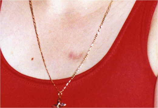
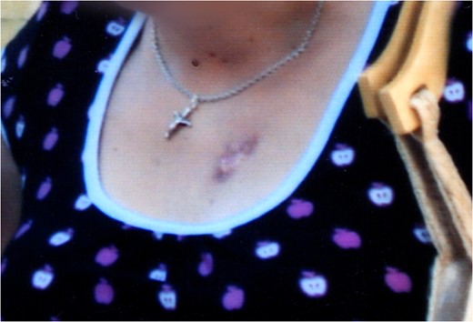
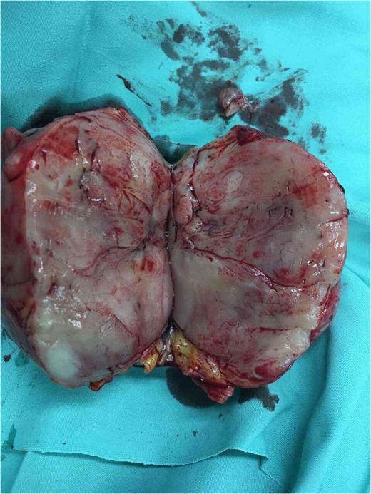
A 48-year-old woman who was born with a vascular malformation in her sternal region (Figs 1 and 2) suddenly noted a mass next to her birthmark.The patient had no significant pain or tenderness. Six months prior to presentation, in our clinic, the patient experienced embolotherapy from a vascular surgeon. As a result, the tumor grew quickly, within a period of 4 months, to a size 10–13 cm (Fig. 3). The patient was subsequently referred to the vascular Unit of Laiko University hospital in Athens. The lesion was seen projecting to the upper anterior chest wall in the sternal region. On physical examination, the mass was huge, quite firm, immobile, with no pain or tenderness in palpation. There were no palpable lymph nodes in the supraclavicular and axillary areas. Other organ examinations were normal. The laboratory studies including complete blood count test, erythrocyte sedimentation rate and C-reactive Proteins were within the normal range.MRI showed a large mass to the upper anterior chest wall most compatible with vascular malformation and there was no evidence of pectoralis major or bone involvement. The patient underwent surgical resection of the tumor (Fig. 4). Histopathology and immunochistochemical staining findings were consistent with the diagnosis of DFSP-FS with close margins and the patient reffered 2 months later to the Sarcoma Unit of Greek Anticancer Institute. We performed a wide local excision with a 3–4 cm margin (Fig. 5) all around the vertical scar in the sternal region. A part of left pectoralis major and the outer layer of the sternal body were also resected. Bilateral, pectoralis major muscle advancement flaps and a full thickness skin graft from the abdominal wall were used to cover the defect resulting from the excision (Figs 6 and 7). Biopsy revealed a residual tumor with a diagnosis of fibrosarcomatous dermatofibrosarcoma (DFSP-FS). Immunohistochemical staining was positive for CD34 (Fig. 8) with rare areas of negative expression. The dermis observed with increased vascularity,a presence of vascular congestion compatible with vascular malformation and a coexistence of scar tissue, suture granulomas and chronic inflammation. The excision margins were clear and radiation therapy was recommended because of the fibrosarcomatous type (Figs 9 and 10).
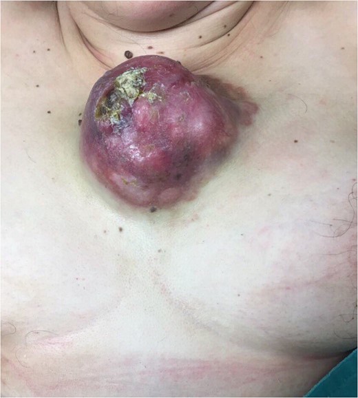
The tumor 6 months prior to presentation in our clinic after embolotherapy.
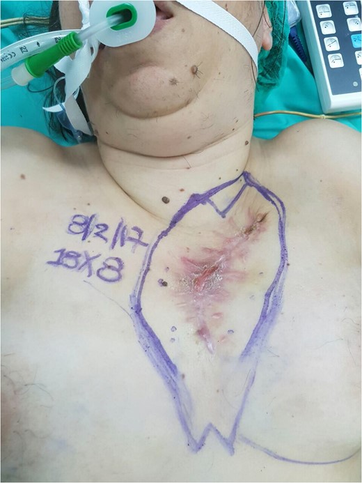
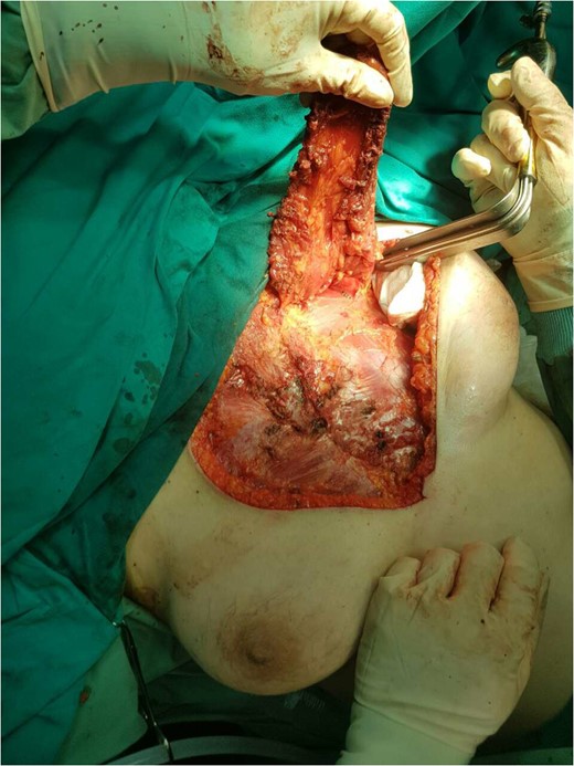
Reconstruction with bilateral pectoralis major muscle advancement flaps.
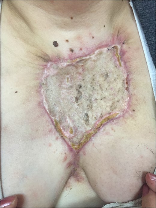
The full thickness skin graft from the abdominal wall to the sternal region 20 days after surgery.
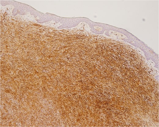
CD34 positive cells. Note the crenz zone between the tumor and epidermis.
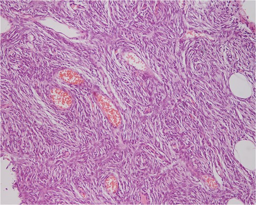
n the wide excision a coincidence of a vascular malformation/carvenous hemangioma in the upper dermal of the neoplasm (DFSP-FS).
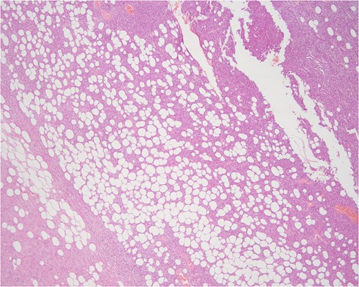
DISCUSSION
There have been several theories on the etiology of DFSP. However, the cause of malignant transformation is as yet unknown. A number of previous reports have suggested a relationship between local trauma such as surgical and old burn scars, previous therapeutic irradiation and sites of vaccinations for DFSP. Fibrosarcomatous DFSP is characterized by an abrupt transition from conventional DFSP to a high grade sarcoma. Typically, it is a fibrosarcoma with the typical herringbone pattern, though rarely a pleomorphic sarcoma (UPS/MFH-like) may be present. The most important feature in the diagnosis is recognizing the precursor DFSP component at the periphery. FS-DFSP is usually positive for CD34, but the fibrosarcomatous component can show reduced expression and, in some cases, loss of CD34 expression. More than 90% of DFSP are characterized by reciprocal chromosomal translocations between chromosomes 17 and 22, t(17;22) or supernumeracy ring chromosomes composed of interspersed sequences from bands 17(17q22) and 22(22q12). It is now clear that this fusion is the main abnormality in its molecular identity. This chromosome rearrangement fuses the strongly expressed collagen 1-Alpha- 1gene (COL1A1) on chromosome 17 with the platelet-derived growth factor (PDGF-B) gene on chromosome 22.PDGF-B, which is a potent mitogen for connective tissue cells, is placed under the control of COL1A1 promoter. This results in autocrine activation of the platelet-derived growth factor receptor tyrosine kinase (PDGF-R), which triggers the proliferation of DFSP tumor cells. FS-DFSP may respond to tyrosine kinase inhibitors like imatinib mesylate for treatment of metastatic or unresectable tumors [1].
A congenital vascular malformation has been known in the past as carvenous hemangioma [2]. PDGF-B has higher levels of expression in carvenous malformations. PDGF-B acts with VEGF on newly formed vessels to specifically to promote stability between the endothelial cells and smooth muscle cells. PDGF pathway, an important signal transfer pathway in angiogenesis, plays a role in the development of vascular malformations and DFSP-FS [3–5]. Malignant transformation in a vascular malformation/hemangioma is very rare and the malignant change has previously been to angiosarcoma [6]. The tumorigenesis in sarcomas is still debated, compared to tumorigenesis in carcinomas where precursor malignancies are well understood. We describe a case of a patient with a presumed vascular malformation in the sternal region for 47 years preceding her diagnosis of DFSP-FS. The possible explanation for this pathologic result is a coincidental DFSP arising at the site of a vascular malformation. PDGF could turn out to be the fundamental intrinsic mechanism of malignant transformation of a vascular malformation. Due to overexpression of PDGF-B an uncontrolled angiogenesis and malignant change to DFSP-FS should be considered. Further studies are needed to determine the exact mechanisms underlying altered PDGF-B expression in the malignant transformation of vascular malformations. In conclusion, our case is the unique case of a DFSP at the sight of a congenital vascular malformation.
CONFLICT OF INTEREST STATEMENT
None declared.
REFERENCES
- radiation therapy
- ulcer
- embolization
- aggressive behavior
- reconstructive surgical procedures
- sarcoma
- soft tissue neoplasms
- sternum
- surgical procedures, operative
- neoplasms
- pathology
- skin cancer
- dermatofibrosarcoma protuberans
- soft tissue sarcomas
- abdominal wall
- full thickness skin graft procedure
- vascular anomalies
- pectoralis major flap
- misdiagnosis
- excision
- wide local excision


