-
PDF
- Split View
-
Views
-
Cite
Cite
Gary Sharp, Daniel Kozman, Inverted Meckel's diverticulum causing intussusception in a Crohn's patient, Journal of Surgical Case Reports, Volume 2015, Issue 9, September 2015, rjv112, https://doi.org/10.1093/jscr/rjv112
Close - Share Icon Share
Abstract
Cases of inverted intraluminal Meckel's diverticulum (MD) containing a lipoma, pancreatic and gastric ectopic tissue resulting in intussusception are extremely rare. We have been unable to locate any such presentation in a Crohn's patient; here, we discuss one such case. MD is the most common congenital malformation of the human gastrointestinal tract. Its paediatric preponderance may result in its oversight in the adult population as a cause for symptoms. Intestinal obstruction is the most common adult presentation in non-inverted MD, whereas haemorrhage and anaemia are the most frequent presenting complaint with an ‘inverted’ MD. Adult intussusception due to an inverted MD is exceptionally rare with a poorly understood pathophysiology and life-threatening consequences. There is no gold standard diagnostic test for an inverted MD causing an intussusception, rather clinicians should utilize many modalities including angiography, ultrasound and computed tomography. Treatment of an inverted adult MD causing intussusception is surgical resection.
INTRODUCTION
Meckel's diverticulum (MD) was first described by Fabricius Hildanus in 1598 and subsequently coined ‘Meckel's diverticulum’ by the German anatomist Johann Friedrich Meckel in 1809 [1]. MD is a true diverticulum resulting from persistence of the vitelline duct [1], which should obliterate by Week 10 of gestation [2, 3]. This small-bowel diverticulum is the most common congenital malformation of the human gastrointestinal tract [1, 2, 4] and is commonly known by the ‘rule of twos’ and found within 2 feet of the ileocaecal junction, affecting 2% of the population, and 2-in. long [1]. In reality, an MD can be found anywhere up to 100 cm proximal to the ileocaecal value on the antimesenteric border [3]; both its diameter and length vary between 1–50 and 1–56 cm, respectively [5, 6]. The majority of MD are found in those <10 years old [6]. Cases of inverted intraluminal MD containing a lipoma, pancreatic and gastric tissue resulting in intussusception are extremely rare. We have been unable to locate any such presentation in a Crohn's patient within available literature.
CASE REPORT
A 30-year-old male, non-smoker, with a known Crohn's disease presented to the emergency department with severe cramping, central abdominal pain and obstipation. He had undergone a right hemicolectomy 2 years prior secondary to an ileocolic intussusception. On examination, his abdomen was tender in the right upper quadrant but not peritonitic. Blood tests, venous lactate and chest radiograph were unremarkable. An intravenous and oral contrast computed tomography (CT) revealed an appearance of intussusception with proximal bowel distention distal to the previous ileocolic resection site.
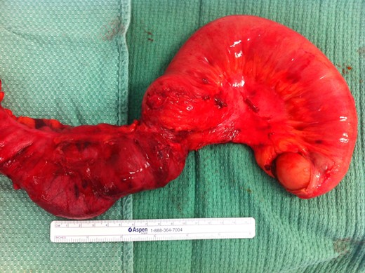
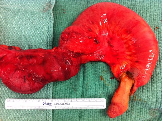
His abdomen was initially explored laparoscopically and then converted to laparotomy due to extensive adhesions. The old ileocolic anastomosis was mobilized and a large polypoid mass was palpable in the small bowel ∼30 cm from the ileocolic anastomosis (Figs 1 and 2). A redo ileocolic side-to-side stapled anastomosis was fashioned. His post-operative recovery was hampered by an ileus, which resolved allowing discharge 7 days after his initial presentation.


Histopathology showed intussusception of an unusual lipoma-like serosal malformation. This included an area of heterotrophic pancreatic tissue with mucosa showing focal gastric type epithelium and glands.
DISCUSSION
The true incidence of MD ranges from 0.6 to 3% [1, 5, 6], with only 4% being symptomatic [2]. Incidence is not affected by gender, but males are more prone to complications [1, 6] and inverted MD (69%) [3]. MD is usually an incidental finding [3] in the paediatric population [1, 6]. Due to this propensity, MD can be overlooked as a potential source of pathology in adults [1] increasing mortality especially in the elderly [6] population.
MD can contain gastric, pancreatic, [3] colonic, jejunal or duodenal ectopic tissue [6] mostly at its tip [1]. Gastric mucosa is the most common in both non-inverted [1] and inverted MD [3]. The lifetime risk of complications associated with an adult MD is 4–16% [1]. Painless haemorrhage is the most common presentation in the ‘paediatric’ population [1, 6] secondary to peptic ulceration from gastric/pancreatic juices [1, 6]. However, intestinal obstruction is the most common adult presentation of MD [1, 4] and can be due to a congenital band from the tail of the MD to the anterior abdominal wall [7] causing a small-bowel volvulus, intussusception or a Littre's hernia [1, 2, 4]. Intussusception is infrequent in adults when compared with children [7], accounting for merely 5% of ‘all’ diagnosed intussusceptions and causing only 1–5% of adult small-bowel obstructions [2, 7]. Adult intussusception due to an inverted MD is exceptionally rare [2].
The pathophysiology of an inverted MD and subsequent intussusception is poorly understood [2–4]. One theory is that the MD base becomes inflamed due to ectopic tissue and these inflammatory changes in turn create abnormal peristaltic contractions resulting in inversion of the MD [2, 4]. The inverted MD then acts as a lead point allowing telescoping of the bowel culminating in an intussusception [2, 4, 7]. Ileocolic intussusceptions are most frequent [7] and result in an oedematous, vascularly compromised bowel wall, which becomes ischaemic and necrotic and ultimately perforates [7]. The median age of those presenting with this rare condition is 27.7 years [3]. Unfortunately, the presentation is broad thus confounding the diagnosis [2], but haemorrhage and anaemia appear to be the most frequent presenting complaint in those with an ‘inverted’ MD [3].
No gold standard diagnostic test exists for an inverted MD causing intussusception. If per-rectal bleeding is the major presenting complaint, then angiography should be utilized if a rate of >0.5 ml/min is expected [1]. Proof of a vitelline artery, a terminating branch of the superior mesenteric artery, on angiography is pathognomonic of an MD [6]. Angiography may even prove to be therapeutic through embolization [6]. If angiography is negative and bleeding remains the main complaint, a red cell scan may be sought [3].
Scintigraphy, first used in 1962 as a diagnostic aid in the MD, is the most common non-invasive modality used [1]. Intravenous Technetium-99m pertechnetate is administered and the isotope is taken up preferentially by the gastric mucosal chloride-secreting cells. In children, it has high sensitivity but low specificity [3]; nonetheless, in adults it is less reliable with greater false negatives and a specificity of only 9% [1]. False positives have also been noted with bowel obstructions, intussusception and intestinal inflammation such as found in Crohn's disease [6].
Some authors suggest that ultrasound should be the first modality used in diagnosing intussusception as it shows the classic ‘target’ or ‘doughnut sign’ [2]. While others advocate CT as the most ‘reliable’ modality to aid preoperative diagnosis and confirm intussusception [2]. In one review, a ‘positive’ CT scan was found in 100% of patents presenting with an inverted MD subsequently found intraoperatively [3]. Images classically show a characteristic ‘sausage-’ or ‘target-’ shaped soft tissue mass [2, 3], or an intraluminal mass with a central area of fat representing the inverted MD and its mesentery surrounded by soft tissue [6].
When a diagnosis of adult intussusception is made, regardless of its cause, surgery is compulsory [3] unless the patient is palliative. Conservative treatment, often utilized to good effect in children, is not appropriate in adults [3].
CONFLICT OF INTEREST STATEMENT
None declared.
REFERENCES
- anemia
- angiogram
- ultrasonography
- congenital abnormality
- computed tomography
- lipoma
- hemorrhage
- crohn's disease
- adult
- ectopic tissue
- diagnostic techniques and procedures
- intestinal obstruction
- intussusception
- pediatrics
- meckel's diverticulum
- pancreas
- gastrointestinal tract
- excision
- gold standard
- chief complaint


