


Abstract
Laparoscopic surgery is the modality of choice in the surgical treatment of colorectal malignancies. The reported incidence of trocar site hernias is 0.65–2.80% with conventional cutting-tip trocars, and this risk increases with the diameter of the trocar. Newer bladeless, blunt-tipped trocars effectively mitigate this risk, and routine fascial closure has been generally deemed unnecessary. We present two cases of trocar site hernias despite the use of bladeless trocars in laparoscopic colorectal surgery, challenging the conventional wisdom of leaving such fascial defects open.
INTRODUCTION
Laparoscopic surgery is increasingly commonplace, and with it, the risk of a trocar site hernia (TSH). Newer ports with bladeless, blunt-tipped trocars are thought to mitigate this risk, making fascial closure of port sites unnecessary. We present two cases of TSH.
CASE REPORT
Our first case is a 72-year-old male who underwent a laparoscopic anterior resection for a rectosigmoid carcinoma. Three ports were used—10-mm port in the umbilicus, 5-mm port in the right lumbar and 12-mm bladeless, blunt-tipped trocar in the right iliac fossa. The resected specimen was retrieved though a 4-cm mini-Pfennestial incision which was closed primarily. A 10F Jackson-Pratt surgical drain was placed in the 12-mm trocar site.
The patient's drain was removed on post-operative day (POD) 4 and was discharged. Later that day, he developed small bowel evisceration through the 12-mm trocar site after a bout of coughing. The eviscerated bowel was gangrenous (Fig. 1) and an emergency resection was performed. The offending fascial defect was sutured close.
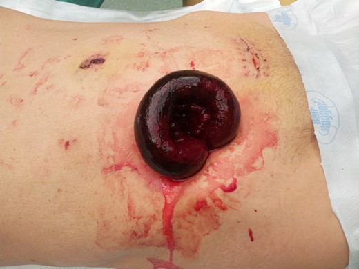
Gangrenous eviscerated small bowel in the 12-mm port site at the right iliac fossa.
Our second case is a 64-year-old female who underwent laparoscopic anterior resection for sigmoid carcinoma in the above fashion. Open fascial closure of the 12-mm port site was performed. No drains were placed. She was discharged on POD 5.
She re-presented on POD 14 with vomiting and abdominal pain. Clinical examination revealed a distended abdomen. A CT scan of the abdomen showed dilated small bowels with a transition point at an ileal herniation through a defect in the right iliac fossa (Fig. 2).
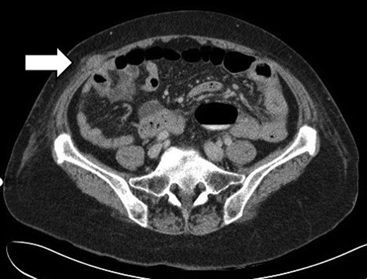
CT scan showing dilated small bowels and a transition point at an ileal herniation through the anterolateral abdominal wall in the right iliac fossa.
The patient underwent emergency surgery. A Richter's type hernia was found at the 12-mm trocar site (Fig. 3). A suture was found in the subcutaneous layer—the fascial-closure stitch at the first surgery might have cut through or caught the wrong layer. The entrapped bowel was viable and reduced successfully. The fascial defect was repaired.
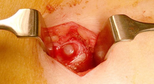
Richter's type hernia in the previous 12-mm port site.
DISCUSSION
Laparoscopic approach is the modality of choice in the surgical treatment of colorectal malignancies, minimizing complications associated with open surgery. It has however its own unique complications, including TSH.
Tonouchi [1] published a review in 2004 where he classified TSH (Fig. 4). The early-onset type involves dehiscence of the anterior and posterior fascial planes, and peritoneum. It develops in the early post-operative period and presents as small bowel obstruction. The late-onset type involves dehiscence of the anterior and posterior fascial planes with an intact peritoneum that forms the hernia sac. This develops months later and may be asymptomatic. The special type represents a full-thickness dehiscence of the entire abdominal wall, with protrusion of the bowel. This presents early, requiring emergency surgery.
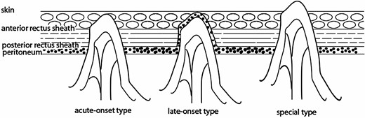
Tonouchi's classification of trocar site hernias.
Tonouchi reported the incidence of TSH to be 0.65–2.80%; the true incidence may be higher as asymptomatic individuals may not seek treatment, and current studies may not report long enough follow-ups.
The risk of TSH increases with trocar size. A survey by the American Association of Gynecologic Laparoscopists showed that 86.3% (725 cases out of 840 TSH) occur with trocar diameters of at least 10 mm. The hernia rate dropped from 8.00 to 0.22% with fascial closure in 12-mm bladed trocars; some authors hence proposed mandatory fascial closure of trocar sites 10 mm or greater [2].
Another factor contributing to TSH is its location. Umbilical sites were the most common due to its anatomical and inherent weakness [3]. Trocar sites situated off-midline were less susceptible due to overlapping of muscle and fascial layers [3]. Lateral abdominal walls composed of two fascial planes and muscles were also less prone to dehiscence [4]. Stretching a port site to retrieve a specimen can extend the fascial defect, predisposing to TSH [5].
Newer ports and bladeless trocars have decreased the incidence of TSH to nearly 0% [6, 7]. These new trocars are atraumatic and split, rather than cut, muscle fibres upon entry. After removal of port, the abdominal layers contract, resulting in a defect much smaller than the trocar diameter [2].
Bhoyrul et al. [6], in his multicentre, randomized trial comparing conventional cutting trocars against bladeless, radially expanding ones, demonstrated that nil fascial closure was safe in the latter, with no TSH after an 18-month follow-up. Other retrospective reviews revealed similar conclusions even for larger bore bladeless trocars [7].
Three studies though have shown TSH with 12-mm bladeless trocars [8–10] in general surgery, although the incidence remains low at 0.40–1.20%. Two [8, 9] of these studies were however heterogenous and included TSH from older cutting trocars, whereas Johnson's [10] TSH incidence of 1.20% all occurred in the umbilicus. There is currently no consensus regarding routine closure of large bladeless trocar sites situated off-midline.
In our study, these two patients represent our first complications of TSH in a personal series of 300 laparoscopic colorectal cases all performed with bladeless trocars; an incidence of 0.6%. One was a special type TSH; the other was an early-onset TSH. Both occurred at the 12-mm trocar sites in the right iliac fossa.
The cause of TSH was likely multifactorial. First, the larger 12-mm trocar created a larger fascial defect. Secondly, the long surgery, port manipulation, and using the port site for drain placement prevented the muscle and fascial planes from overlapping. Finally, placing the trocars in the right iliac fossae, which lacked a posterior fascial plane, further promoted TSH.
One patient had a coughing fit, and the second was obese—both patient factors for TSH. Despite attempts at fascial closure of the trocar site in the second patient, a TSH developed, highlighting the need for proper closure of the correct layer under direct vision.
In conclusion, the presentation of a TSH can be overt (special type TSH) or insidious (early-onset type Richter's hernia), and early identification is necessary for good patient outcome. Recognizing the contributing factors helps minimize its occurrence.
Bladeless trocars, previously thought to negate the need for fascial closure, have been implicated in TSH formation, challenging the wisdom of leaving such fascial defects open. We feel that every effort should be made to close the defect. Direct visualization of the fascia is critical for proper closure; the use of S-retractors for visualization in open closure, or the use of suture-passers in laparoscopic-assisted closure may be helpful.
FUNDING
No financial grants or funding were provided. The authors do not have any industrial links or affiliations.
CONFLICT OF INTEREST
All authors have no conflict of interest.
