


Abstract
Femoral neck fractures in young patients can have unusual presentation making diagnosis and subsequent management more difficult. Early detection is essential to avoid complications; however, it is only achievable with a high index of suspicion. We present an unusual case of a 39-year-old office worker who presented with a 5-month history of spontaneous onset of hip pain. She had multiple clinic visits prior to the diagnosis of neck of femur fracture. Fracture displacement and delayed diagnosis had resulted in a non-union by the time of presentation. She was subsequently managed with a fixation using a dynamic hip screw along with subtrochanteric osteotomy. No underlying cause for the initial fracture was identified despite thorough investigation. Learning points for early detection and operative management are discussed.
INTRODUCTION
Femoral neck fractures in young populations usually result from high energy trauma [1]. Non-traumatic femoral neck fractures have also been reported in young athletes and military personnel due to stress fractures [2, 3]. Delayed diagnosis in such populations is high, with time to diagnosis reported to be 14 weeks on average [4] and in some cases considerably longer. The delay is usually due to a low index of suspicion combined with nebulous symptoms. Complications resulting from delayed treatment have significant impact on patients with continued pain and impaired mobility. The risk of fracture displacement also increases with delayed diagnosis, bringing with it the increased risk of non-union and avascular necrosis (AVN). An acutely displaced femoral neck fracture carries a risk of non-union of around 33% and AVN 28% [5]. In this case report, we present a young patient with an unusual presentation of femoral neck fracture and delayed diagnosis.
CASE REPORT
A 39-year-old office worker presented to the fracture clinic with 5 months of right groin pain and difficulty in mobilization. She reported spontaneous, acute onset of pain with little improvement with analgesia and sufficient intensity to prevent weight-bearing. Six months prior to the onset of symptoms, she recalled slipping on a wet floor. However, she did not have pain until 6 months later and had not associated this event with her current problems. She was previously fit and active taking only a contraceptive pill as regular medication. She had no risk factors for stress fractures. Prior to review in the fracture clinic, she consulted a number of health-care professionals on multiple occasions. She was initially diagnosed with sciatica and given analgesia and physiotherapy. With little improvement in her symptoms and repeated visits to the emergency department and her general practitioner, she was suspected to have trochanteric bursitis and given a steroid injection and further physiotherapy. Throughout this period her symptoms remained unchanged and she required a stick for mobilization. Five months later, an X-ray was finally requested revealing a fracture and prompting referral to the fracture clinic.
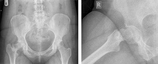
Pelvic and right hip radiograph's taken 5 months after the onset of symptoms demonstrating the fracture neck of femur.
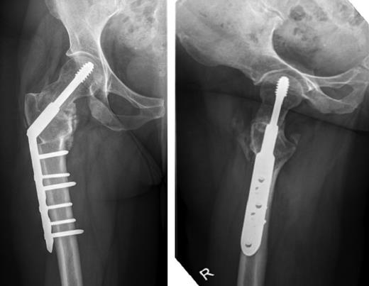
Anterior-posterior and lateral radiographs taken 6 months postoperatively demonstrating union of the fracture and osteotomy site.
DISCUSSION
Morbidity from femoral neck fractures is linked to the degree of fracture displacement and risk of AVN and non-union [2]. Other complications include disuse osteopenia, femoral neck resorption, proximal migration of the greater trochanter, difficult reduction and high failure rates of internal fixation [6]. Management options for missed femoral neck fractures, broadly speaking, are either femoral head replacement, or salvage surgery with some form of fixation. In young patients, preservation of the femoral head is preferable with joint replacement reserved as a last resort. A number of fixation methods have been used with varying success [7]. Valgus intertrochanteric osteotomy, where a lateral wedge is removed at the upper border of the lesser trochanter, allows lateral displacement of the distal fragment and valgus correction of the deformity. This has been shown to be successful in treating missed femoral neck fractures with favourable outcomes [3, 6 –8]. However, under-correction of the deformity can lead to failure of the fixation [7] and subsequent conversion to hip replacement will technically be more difficult as a secondary procedure [8].
Non-union from femoral neck fractures is a result of both mechanical and biological factors and both must be addressed to allow union. Valgus osteotomy addresses mechanical factors by correcting the deformity and stabilizing the fracture. The vascular supply to the femoral head must also be considered. Closed reduction should be achieved where possible to preserve the retinacular vessels and some advocate using a limited antero-lateral approach to enter the hip (as was used in this case) to ensure preservation of the blood supply. This also allows easy removal of fibrous tissue from the fracture site to encourage re-vascularization [6].
This case emphasizes a number of learning points. It was an unusual presentation of femoral neck fracture. Numerous opportunities to diagnose the fracture were missed and the diagnosis was delayed for 5 months. Following her initial diagnosis of sciatica, she was reviewed by a further three health-care professionals. All continued to treat for sciatica without considering an alternative diagnosis until her fifth attendance when a diagnosis of trochanteric bursitis was made, prompting steroid injections and further physiotherapy. This also failed to improve her symptoms and by this time, her mobility had significantly deteriorated. No X-rays were taken in any of these visits. This emphasizes the importance of re-assessing patients and determining whether treatment has had the expected result, and if not, to consider alternative diagnoses and further investigations.
During this 5-month period the fracture displaced, with shortening of the femur and varus deformity of the hip. Clinically, this resulted in a leg-length discrepancy of 4 cm and significant abductor weakness with the positive Trendelenburg test. The patient required a walking stick for mobilization and struggled to continue working. These clinical findings were easily elicited from orthopaedic examination and should have alerted the clinician to the diagnosis.
Finally, no underlying cause for the fracture was found despite thorough investigation. Although there was a history of trauma, this was 6 months prior to the onset of symptoms. No underlying bone pathology was detected on MRI scan and blood tests were all normal. The patient was not an athlete and had no other risk factors for a stress fracture.
In summary, delayed diagnosis of femoral neck fractures in young patients is difficult to manage and early detection is essential to prevent fracture displacement and the subsequent associated risks of non-union and AVN. However, diagnosis can only be achieved if clinicians have a high index of suspicion and request appropriate investigation following a detailed history and clinical examination.
CONFLICT OF INTEREST STATEMENT
None declared.
