-
PDF
- Split View
-
Views
-
Cite
Cite
D Henry, S Satgunam, Idiopathic omental bleeding, Journal of Surgical Case Reports, Volume 2012, Issue 9, September 2012, Page 2, https://doi.org/10.1093/jscr/2012.9.2
Close - Share Icon Share
Abstract
Omental bleeding without any evidence of trauma, aneurysms, or other pathology has been rarely described in the literature. We report a case of a 24 year-old female on aspirin/acetaminophen/caffeine for migraines who presented with abdominal pain and tachycardia. Computed tomography angiography revealed active extravasation in the right lower quadrant. During exploratory laparotomy, a small bleeding artery within the greater omentum was suture ligated, and two liters of fresh and clotted blood were evacuated. The patient recovered successfully. We review the diagnosis and management of this rare condition.
INTRODUCTION
The first known report of spontaneous bleeding from the greater omentum occurred at Montreal General Hospital in 1918 (1). Since then, omental bleeding has been associated with trauma, neoplasms, omental torsion (2), varices, aneurysms (4,5), and vasculitis (6). Hemoperitoneum is common, however, idiopathic bleeding from the greater omentum is a rare event (1,7,8). We discuss a case of hemoperitoneum due to bleeding from a small omental artery, which was treated successfully with laparotomy and suture ligation.
CASE REPORT
A 24 year-old, obese, Caucasian female presented to an outside hospital complaining of malaise, myalgias, and fatigue. Viral myalgia was diagnosed, and she was discharged home. Three days later, she presented to the emergency department complaining of diffuse abdominal pain, which began when she vomited and had an episode of syncope that morning. A computed tomography (CT) scan of the brain was negative. A CT scan of the abdomen and pelvis revealed diffuse free fluid, consistent with hemoperitoneum (41 Hounsfield units). She suffered physical abuse a year ago but denied any recent abuse or other trauma.
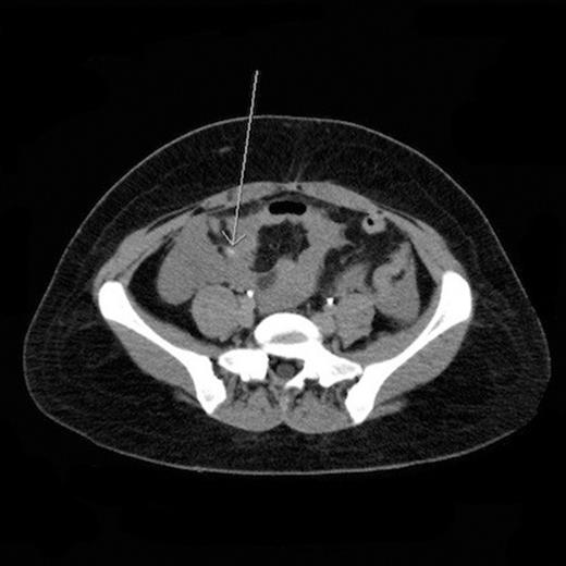
A computed tomography angiogram (CTA) was performed with arterial and venous phase contrast, and active arterial extravasation along the mesentery within the right lower quadrant was identified (Fig. 1). The spleen was also mildly enlarged at 15 cm in length. At that time, her temperature was 99.2°F, heart rate was 110-120 bpm, respiratory rate was 20/minute, blood pressure was 90/67 mmHg, and oxygen saturation was 98% on two liters/minute nasal cannula oxygen. Pertinent laboratory values are as follows: white blood cell count 8.3 x 10³/µL, hemoglobin 9.8 g/dL, hematocrit 29.7%, platelets 135 x 103/µL, creatinine 0.91 mg/dL, INR 1.1, and lactic acid 2.7 mmol/L.

CT angiogram of the abdomen and pelvis revealed active extravasation within the right lower quadrant
Her past medical history was significant for migraines and four Cesarean sections. Her only home medication was aspirin/acetaminophen/caffeine.
Intravenous Lactated Ringer’s solution, two units of packed red blood cells, morphine, a small dose of lorazepam for anxiety, and tranexamic acid were administered. After these interventions, the patient’s heart rate was 86 bpm, and hemoglobin was 8.0 g/dL. During this time, the patient refused to undergo surgery. Once she consented, an emergent laparotomy was performed.
Upon opening the abdomen, a tiny artery within the greater omentum with active, pulsatile bleeding was ligated with 3-0 silk suture. Hemostasis was achieved. The omentum was otherwise normal. Two liters of fresh and clotted blood were evacuated from the peritoneal cavity. The abdomen was packed and irrigated twice. The organs and mesentery were inspected, and no other bleeding was identified. Few pelvic adhesions, due to her prior Cesarean sections were identified and taken down. Intraoperatively, she received two units of packed red blood cells and two units of platelets, to counteract the effects of aspirin. She recovered well and was discharged home on postoperative day six.
DISCUSSION
Omental bleeding is known to occur after penetrating or blunt trauma, including high-speed deceleration events in the presence of intraabdominal adhesions. Omental bleeding has also been associated with varices (3), hemangiopericytomas, leiomyoblastomas, omental torsion (2), aneurysms (4), and an aneurysm in a peritoneal dialysis patient (5), none of which were identified in the described patient. Generalized pathologic conditions associated with spontaneous bleeding have included segmental arterial mediolysis (segmental mediolytic arteritis), systemic lupus erythematosus, Ehlers-Danlos syndrome type IV (9), fibromuscular dysplasia (9), hypertension, polycythemia vera, and Wegener’s granulomatosis (6).
Coagulopathy due to anticoagulants and antiplatelet agents has also contributed to episodes of spontaneous intra-abdominal hemorrhage. Finally, Finley (2005) reported a mortality from the rupture of an omental varix in a patient taking Sildenafil citrate. Our patient’s last dose of Aspirin/Acetaminophen/Caffeine was the night before her hospital presentation, and the platelet function analysis was abnormal. Thus, platelet dysfunction likely contributed to her persistent, active bleeding from a tiny artery.
She denied any recent trauma, however, she had forceful vomiting on the morning of her hospitalization. She also had adhesions due to her four Cesarean sections. A postulated mechanism for the patient’s omental bleed is that forceful vomiting caused avulsion of an omental adhesion (8), causing bleeding. Abuse or trauma that the patient failed to admit are also possible. However, no visual or radiographic evidence of soft tissue contusions or solid organ injuries were present.
The workup and management of spontaneous intra-abdominal hemorrhage, without any antecedent trauma, should begin with the stabilization of the patient’s airway, breathing, and circulation. Coagulation studies (7) and possibly a platelet function analysis should be considered. And reversal of coagulopathy may include platelets, fresh frozen plasma, vitamin K, factor IX complex, and/or tranexamic acid.
Imaging and diagnostic studies useful in the case of spontaneous intra-abdominal hemorrhage include contrast-enhanced CT (4,6-10), CTA (1,4,10) (with arterial or arterial and venous phase contrast), ultrasonography (4,7,9,10) (less useful), diagnostic peritoneal lavage (7), and conventional angiography (7,9,10). Ascitic fluid may be differentiated from blood on CT on the basis of Hounsfield units (HU) and other radiographic characteristics (water: 0 HU; acute blood: 30-45 HU; clotted blood: 50-60 HU; extravasating blood: >85 HU). Many case reports of omental bleeding mention preoperative suspected diagnoses of appendicitis and peritonitis (2,8). Measuring Hounsfield units may expedite the diagnosis and treatment of hemoperitoneum.
If hemorrhage is thought to be massive or ongoing, the patient should proceed emergently to the operating room (2,5-7) or angiography suite (9,10). The greater omentum is supplied by an arcade of collateral arteries branching off of the left and right gastroepiploic arteries (9). The left gastroepiploic artery branches off of the splenic artery. The right gastroepiploic artery is one of two terminal branches of the gastroduodenal artery, the other of which is the anterior superior pancreaticoduodenal artery. Matsumoto (2011) reported angiographic embolization of the left gastroduodenal artery in a patient with a similar presentation of spontaneous bleeding from an omental artery. This successful acute treatment was then followed 10 days later by a partial omentectomy, due to a suspicion for neoplasia.
Open surgical technique includes suture ligation or resection for omental bleeding, clot evacuation, irrigation, and four-quadrant packing if the source is not initially identified. Inspection of the spleen and other organs should be performed, as well as the mesentery and omentum. In the presented case, suture ligation of the bleeding tiny omental artery was performed. However, in almost every other reported case of omental bleeding, a partial omentectomy was performed (2,4-9).
In conclusion, idiopathic bleeding from the greater omentum is a rare cause of potentially life-threatening hemoperitoneum and hemorrhagic shock (5,7). Many patients have a history of abdominal surgery, and a torn omental adhesion is a possible cause of many cases of otherwise idiopathic omental bleeding. On CT scans, ascitic free fluid should be differentiated from blood (8) on the basis of Hounsfield units. Treatment consists of emergent laparotomy or angiographic embolization.


