


 https://orcid.org/0000-0002-7096-9736

Abstract
Ectopic teeth are rare entities occurring outside their normal anatomical position, and ectopic mandibular premolars are particularly uncommon. This report describes the management of a deeply ectopic mandibular second premolar located at the inferior border of the mandible. A 16-year-old male patient was referred following incidental radiographic detection. Cone-beam computed tomography revealed buccal cortical perforation and a radiolucent lesion suggestive of a dentigerous cyst. Due to the deep location, limited intraoral access, and proximity to the inferior alveolar nerve, the tooth was removed under general anesthesia using an extraoral submandibular approach. Histopathological examination confirmed a dentigerous cyst. The postoperative course was uneventful, and complete bone healing was observed at 1-year follow-up. The extraoral approach may represent a safe and effective option in selected cases with limited intraoral accessibility.
Introduction
Ectopic teeth are rare clinical entities that develop outside their normal anatomical position and are often detected incidentally during routine radiographic examination. The reported prevalence ranges between 0.1% and 1% in the general population [1]. They are most commonly located in the maxillary sinus and palatal region, whereas involvement of atypical sites such as the nasal cavity, condyle, coronoid process, and orbit is less frequent [2]. The majority of cases involve the mandibular third molars, while the premolars are rarely affected [3].
The etiology of ectopic eruption is multifactorial, including trauma, infection, developmental disturbances, and odontogenic pathology. Dentigerous cysts may contribute to tooth germ displacement, resulting in ectopic positioning [3]. In addition, disruption of epithelial–mesenchymal interactions during odontogenesis has been suggested [4].
Management depends on the anatomical location, symptoms, and associated pathology. Surgical removal is indicated in cases with cystic changes or a risk of complications [1]. The choice of surgical approach is determined by the three-dimensional position of the tooth and its relationship with adjacent structures. Although intraoral approaches are generally preferred, extraoral approaches may be required for deeply located cases [5].
Case report
A 16-year-old male patient was referred following the incidental detection of an ectopic mandibular second premolar near the inferior border of the left mandible (Fig. 1). The patient was asymptomatic and systemically healthy. The dental history revealed early extraction of the permanent first molar in the same region.
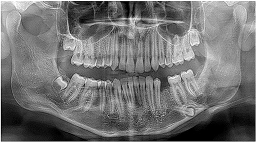
Preoperative panoramic radiograph showing an ectopic mandibular second premolar near the inferior border of the left mandible.
Cone-beam computed tomography (CBCT) revealed the precise three-dimensional position of the ectopic tooth near the inferior cortical border of the mandible, with buccal cortical perforation and an associated radiolucent lesion suggestive of a dentigerous cyst (Figs 2 and 3). This imaging was critical for surgical planning and for selecting the extraoral approach.
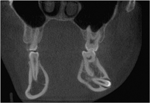
Coronal CBCT image demonstrating that the ectopic premolar was positioned close to the inferior cortical border of the mandible, with buccal cortical perforation and associated follicular enlargement.
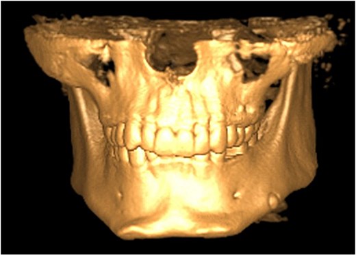
Three-dimensional reconstruction showing buccal cortical perforation associated with the ectopic tooth and its inferior migration within the mandibular body.
Owing to the deep location, limited intraoral access, proximity to the inferior alveolar nerve, and risk of mandibular fracture, an extraoral approach was planned. Surgery was performed under general anesthesia. A submandibular incision was made, followed by careful dissection of the platysma. The marginal mandibular branch of the facial nerve was identified and preserved during surgery. Access to the mandible was achieved, and the existing cortical perforation was used to facilitate the visualization. The tooth and associated pathological tissues were atraumatically removed (Fig. 4). The surgical site was irrigated and closed in a layered manner.
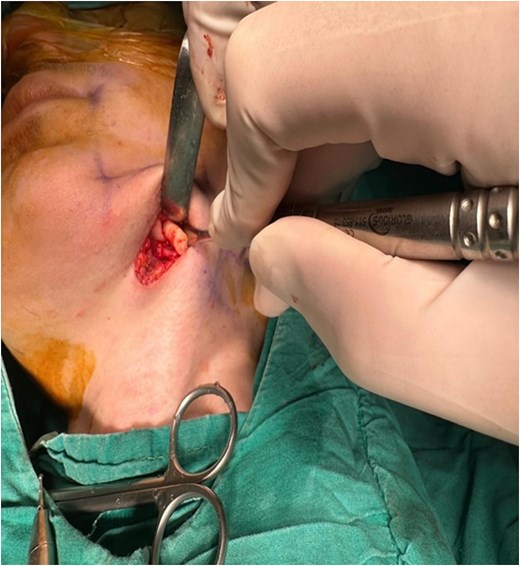
Intraoperative view during the submandibular approach demonstrating direct surgical access to the ectopic premolar following enlargement of the existing cortical perforation.
Histopathological examination confirmed the diagnosis of a dentigerous cyst. The postoperative course was uneventful, with no infection, hematoma, or neurosensory deficits. At the 1-year follow-up, complete bone healing was observed, and the extraoral scar was esthetically acceptable (Fig. 5).
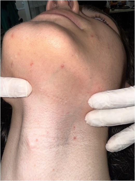
Extraoral view at the 1-year follow-up showing satisfactory healing at the submandibular incision site with minimal scar formation.
Discussion
Ectopic mandibular premolars are rare, and only a limited number of cases have been reported in the literature [3, 6–8]. Previous reports have also indicated that intraosseous migration of mandibular second premolars is uncommon, with bilateral migration being considerably rarer than unilateral migration.
The exact etiology of ectopic eruption remains unclear; however, several contributing factors have been proposed, including trauma, infection, developmental disturbances, and odontogenic pathology. Among these, dentigerous cysts are considered one of the most significant factors, as cystic expansion may displace the developing tooth germ and lead to ectopic positioning [3]. In the present case, histopathological confirmation of a dentigerous cyst supports this mechanism. Additionally, a history of early extraction of the permanent first molar in the same region may have contributed to abnormal eruption patterns and distal migration of the premolar, as suggested in previous studies [9].
The clinical management of ectopic teeth depends on multiple factors, including the anatomical location, presence of symptoms, and associated pathological findings. In asymptomatic cases without evidence of pathology, a conservative approach with periodic clinical and radiographic follow-ups may be considered [1]. However, surgical intervention is indicated in the presence of cystic lesions, cortical perforation, infection risk, or potential complications affecting adjacent structures of the jaw. In the present case, the presence of significant follicular enlargement and buccal cortical perforation strongly indicated the need for surgical intervention.
Accurate preoperative evaluation plays a critical role in determining the appropriate surgical approaches. CBCT provides detailed three-dimensional information regarding the exact localization of ectopic teeth and their relationships with surrounding anatomical structures [10, 11]. In this case, CBCT imaging was essential for identifying the inferior position of the tooth, its proximity to the inferior alveolar nerve, and the presence of cortical perforation. This information is crucial for minimizing intraoperative risks and selecting the most appropriate surgical technique.
The choice of surgical approach remains a key consideration in the management of deeply impacted teeth. Although intraoral approaches are generally preferred because of their minimally invasive nature and absence of external scarring, they may be inadequate in cases involving deep anatomical localization or limited surgical access. In such situations, the intraoral approach may require extensive bone removal and increase the risk of injury to vital structures, particularly the inferior alveolar nerve. Conversely, extraoral approaches provide improved visualization, direct access, and greater control during the procedure, thereby enhancing the surgical safety [5]. Previous studies have reported the successful use of extraoral approaches in challenging anatomical regions, including the inferior border of the mandible, condyle, coronoid process, and ramus [12, 13]. Despite these advantages, the extraoral approach is associated with potential complications, such as facial nerve injury and postoperative scar formation. Therefore, careful patient selection and meticulous surgical techniques are essential to minimize these risks. In the present case, the identification and preservation of the marginal mandibular branch of the facial nerve, along with the use of an existing cortical perforation, allowed for the safe and effective removal of the ectopic tooth without complications. Overall, this case highlights that in deeply positioned and anatomically complex ectopic mandibular teeth, particularly in the presence of cortical perforation and limited intraoral accessibility, the extraoral approach can be considered a reliable and effective surgical alternative when supported by thorough preoperative planning and appropriate patient selection.
Conclusion
Deeply ectopic mandibular second premolars require careful evaluation and individualized treatment planning. When intraoral access is limited, the extraoral submandibular approach is a safe and effective alternative.
Conflicts of interest
The authors declare no conflict of interest.
Funding
No funding to declare.
Ethics
Ethical approval was not required for this case report.
Consent
Written informed consent was obtained from the patient.


