


 https://orcid.org/0000-0001-7699-3342

Abstract
Metastasis of oropharyngeal squamous cell carcinoma (OPSCC) to the thyroid gland is rare, and p16-positive cases form an even smaller subset. We present a case of p16-positive thyroid metastasis from OPSCC identified after complete treatment response. Nine months after completing chemoradiotherapy, the patient developed throat pain and hoarseness. Computed tomography demonstrated an enlarged heterogeneous right thyroid gland and fine needle aspiration confirmed p16-positive metastatic OPSCC in the absence of locoregional recurrence. The patient underwent extended total thyroidectomy and right selective neck dissection; formal pathology confirmed metastatic OPSCC with involved margins and 13/22 positive lymph nodes. To our knowledge, this is the first reported intrathyroidal metastasis from p16-positive OPSCC after complete treatment response and in the absence of local recurrence. Further cases are required to better understand the epidemiology, optimal management, and prognosis of this presentation.
Introduction
The incidence of oropharyngeal squamous cell carcinoma (OPSCC) is expected to increase 30% by 2030 [1, 2]. HPV-positive OPSCC accounts for up to 80% of OPSCCs in some regions [3]. OPSCC frequently metastasises via the lymphatic route and those from the base of tongue may have bilateral metastases [4, 5], although distant metastases are rare [6]. The most common site of distant metastasis is the lungs, followed by bones, liver, and skin [7]. Intrathyroidal metastasis is very rare, with only a small number of cases reported in the literature, and an even smaller number positive for p16 overexpression [7, 8]. The treatment and prognostic outcomes of this small group of patients remain unclear, especially in patients who have already received curative treatment for their primary disease. Here, we present the case of a patient who developed a p16-positive thyroid metastasis after primary chemoradiotherapy for OPSCC.
Case report
A 55-year-old man presented with a 1-month history of haemoptysis and right-sided ear pain. He had no significant medical history and was a lifelong non-smoker. Examination and computed tomography (CT) revealed a right tonsillar mass and multiple necrotic lymph nodes in the neck. Fine needle aspiration (FNA) demonstrated OPSCC but p16 status was inconclusive. Subsequent tissue biopsy demonstrated moderately differentiated OPSCC, positive for p16 overexpression and positive for p40. Definitive chemoradiotherapy including three cycles of cisplatin and 70 Gy of fractionated radiation achieved a complete treatment response on positron emission tomography-CT (PET-CT) 3 months after treatment (Fig. 1). Two subcentimetre lymph nodes at levels III and IV remained on the left, but follow-up ultrasound demonstrated no concerning features and both regressed completely.
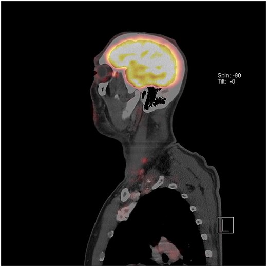
Sagittal PET-CT scan 3 months after definitive chemoradiotherapy for p16-positive OPSCC. Complete treatment response was observed at the primary site (not shown) and the two visible lymph nodes at levels III and IV regressed with follow-up.
At routine follow-up 9 months later, the patient complained of new throat pain and hoarseness. CT demonstrated an enlarged and heterogeneous right thyroid gland with a 36 mm intrathyroid mass invading trachea and oesophagus accompanied by enlarged right paratracheal lymph nodes. PET-CT revealed uptake in those areas (Fig. 2), with no evidence of locoregional recurrence. US-guided FNA of the thyroid mass demonstrated keratinization, no lymphoid cells, and immunohistochemistry was strongly positive for p16. Therefore the thyroid mass was thought more likely to represent metastatic disease than anaplastic thyroid carcinoma with complete squamous differentiation at a multidisciplinary meeting, and was assigned a TNM stage of rT0 rN0 rM1, which maps to rStage IV. The patient was offered and underwent extended total thyroidectomy, partial tracheal and oesophageal resection with primary anastomosis and closure, right selective neck dissection of levels II to VI, and a pectoralis major flap reconstruction. Tumour was found to be invading the trachea and oesophagus and intraoperative frozen sections demonstrated satellite tumours adjacent to the anterior margin. The decision was made not to proceed to a total laryngectomy due to morbidity and low chance of cure. Formal pathology of the 55 × 31 × 63 mm specimen demonstrated metastatic OPSCC in the thyroid with positive margins into the trachea, oesophagus, inferior cricoid, and right recurrent laryngeal nerve and 13/22 lymph nodes were positive with extensive extracapsular spread. The patient declined palliative radiotherapy and subsequently died.
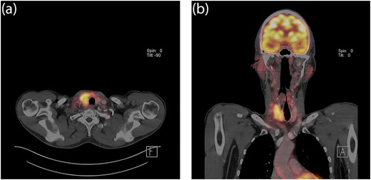
Axial (a) and coronal (b) views of PET-CT scan 9 months after definitive chemoradiotherapy demonstrating avid disease in the right thyroid gland and adjacent paratracheal lymph nodes.
Discussion
Metastatic thyroid disease is rare, with the overall incidence of metastasis of any primary malignancy to the thyroid gland representing only 2%–3% of thyroid gland malignancies [9]. Autopsy series most commonly identify the lungs as the site of primary malignancy in cases of thyroid metastasis [10], whereas renal cell carcinoma is more common in clinical series [10, 11]. OPSCC metastasis to the thyroid gland is rare, with only a small number of published cases, and p16-positive metastases to the thyroid gland are rarer still [11]. Four p16-positive OPSCC metastases to the thyroid gland have been documented in the past [7, 8, 11, 12], but we believe this to be the first published case after complete treatment response and in the absence of local recurrence.
These five cases including the one presented here share some demographic features but demonstrate substantial clinical heterogeneity (Table 1). The timing of thyroid metastasis varied widely. In two cases, the thyroid lesion was diagnosed synchronously with the oropharyngeal primary [8, 11], suggesting either advanced disease at presentation or unusually early haematogenous dissemination. In another case, thyroid metastasis was identified 4 years after the primary diagnosis but only in the context of local recurrence [7]. Our patient is unique in this small cohort in having developed isolated thyroid metastasis 9 months after primary treatment, in the absence of local recurrence. This presentation is more difficult to reconcile with continuity of locoregional disease and may reflect distant haematogenous spread, although the burden of nodal disease (13 of 22 involved nodes) means retrograde lymphatic dissemination cannot be excluded.
Comparison of identified published cases of p16-positive OPSCC metastasis to the thyroid
| Case [ref.] | Age, sex | OPSCC location | Primary treatment of OPSCC | Time interval (OPSCC diagnosis to thyroid met.) | Presenting symptoms of thyroid met. | Salvage therapy for thyroid met. | Treatment outcome |
|---|---|---|---|---|---|---|---|
| 1 [12] | 69, male | Pharyngeal | Chemoradiotherapy | 36 months | Dysphagia, hoarseness | Supportive care | Patient died |
| 2 [7] | 58, male | Tonsillar | Chemoradiotherapy | 48 months | Dysphagia | Chemotherapy | Not reported |
| 3 [8] | 68, male | Tonsillar | Chemotherapy, chemoradiotherapy | 0 months | Neck mass | Total thyroidectomy | MRI at 6 months showed no recurrence |
| 4 [11] | 55, male | Tonsillar | Chemotherapy | 0 months | Neck mass | Total thyroidectomy | Patient remained alive but with disease at 15 months |
| Present case | 55, male | Tonsillar | Chemoradiotherapy | 9 months | Throat pain, hoarseness | Extended total thyroidectomy | Patient died |
Presenting features and management were similarly varied across the cases. Symptoms reflected the location of the thyroid lesion or its mass effect (neck mass [8, 11], dysphagia [7, 12], and hoarseness [12]), rather than features specific to metastatic disease, underscoring the diagnostic challenge in distinguishing metastatic OPSCC from primary thyroid pathology on clinical grounds alone. Primary management approaches were chemoradiotherapy with or without additional rounds of chemotherapy [7, 8, 12]. Salvage therapy ranged from total thyroidectomy with or without follow-up rounds of chemotherapy [8, 11], only chemotherapy [7], or supportive care [12]. Extended total thyroidectomy was offered and performed for the patient presented here. This variability reflects the absence of established guidance for this rare presentation and highlights the difficulty of extrapolating management decisions from such a small evidence base.
Distinguishing metastatic OPSCC from a primary thyroid malignancy or direct local invasion is not always straightforward and requires careful integration of imaging, cytology, and immunohistochemistry [13]. FNA is not always reliable at differentiating between primary and secondary disease [13], but detecting p16 overexpression helped determine the likely oropharyngeal origin of the tumour in this case, as anaplastic thyroid carcinoma has been known to show squamous differentiation [14]. Furthermore, although PET-CT has a good specificity and high negative predictive value for recurrent or persistent head and neck malignancy, there is some evidence that it is less sensitive when performed < 10 weeks after treatment [15]. This case reinforces the value of p16 immunostaining as part of the diagnostic workup when squamous malignancy is identified in the thyroid gland in patients with a prior OPSCC history, particularly where complete treatment response has been documented.
We present a case of p16-positive thyroid metastasis from OPSCC occurring after confirmed complete treatment response and in the absence of locoregional recurrence. This case highlights that thyroid metastasis should remain on the differential even in patients who have achieved complete response to curative-intent chemoradiotherapy. Further cases are needed to better characterize the epidemiology, optimal management, and prognosis of this uncommon pattern of disease spread.
Acknowledgements
W.H.C. is supported by a Woolf Fisher Scholarship from the Woolf Fisher Trust of New Zealand and Cambridge Commonwealth, European & International Trust, and by Christ’s College, Cambridge.
Conflicts of interest
The authors have no conflicts to declare.
Funding
This study did not receive any funding or financial support.
Data availability
Not applicable as this is a case report.
Patient consent statement
Not applicable as we are not publishing identifying digital images or photographs.


