


 https://orcid.org/0000-0002-0505-6009

Abstract
Perihilar cholangiocarcinoma (pCCA), originating from the second-order bile ducts and extending to the cystic duct insertion, is the most common cholangiocarcinoma subtype and carries a dismal prognosis. It is typically characterized by obstructive jaundice and elevated serum carbohydrate antigen 19-9 (CA19-9); however, these features significantly overlap with those of benign biliary diseases. While endoscopic retrograde cholangiopancreatography-guided tissue sampling is crucial for diagnosis, its sensitivity remains suboptimal. Consequently, up to 15% of preoperatively suspected pCCA cases are confirmed as benign strictures postoperatively. Given the close association between choledocholithiasis and pCCA, distinguishing malignancy from stone-induced inflammatory disease remains a significant diagnostic challenge. Herein, we report a rare case of chronic cholangitis mimicking pCCA, ultimately attributed to calculi containing a retained foreign body.
Introduction
Perihilar cholangiocarcinoma (pCCA), classically defined by Altemeier (1957) and Klatskin (1965), arises at the biliary confluence or the proximal right and left hepatic ducts [1]. It is the most prevalent cholangiocarcinoma (CCA) subtype, accounting for ~50%–70% of cases [1], and carries a dismal prognosis [2]. Although precise incidence data are often obscured by historical misclassification as intrahepatic cholangiocarcinoma, its prevalence is notably high in East Asia (e.g. Thailand and China), largely driven by liver fluke infestations [3]. Chronic calculous cholangitis is another well-established risk factor, driven by persistent inflammation and biliary epithelial injury [4, 5]. Despite advances in diagnostic imaging and endoscopy, distinguishing chronic cholangitis from pCCA remains a formidable challenge due to overlapping clinical, biochemical, and radiological features. Herein, we report a rare case of chronic cholangitis secondary to bile duct calculi containing a retained foreign body, which mimicked pCCA.
Case report
A 74-year-old female was admitted following the identification of a space-occupying lesion in the proximal common bile duct (CBD) on computed tomography (CT) at an outside hospital 1 week prior. The patient had a history of deaf-blindness since childhood. Her medical history was significant for prior pulmonary and thyroid surgeries (details unavailable), a cholecystectomy 3 years prior, and a cerebral infarction 3 months before admission.
Upon admission, the patient presented with no fever or jaundice. Carbohydrate antigen 19–9 (CA19–9) was elevated to 255.6 U/ml. Magnetic resonance imaging (MRI) revealed hilar bile duct dilation, stones, and wall thickening suggestive of field cancerization (Fig. 1A–C), along with a renal mass suspicious for renal cell carcinoma (Fig. 1D). Initial endoscopic retrograde cholangiopancreatography (ERCP) visualized a giant stone (Fig. 1E), prompting the placement of a biliary plastic stent (Fig. 1F). Brush cytology was unsuccessful as the brush could not traverse the impacted stone.
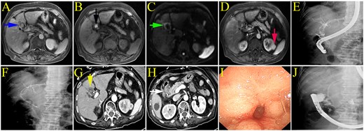
(A–C) MRI demonstrated hilar bile duct dilation (blue arrow), hilar bile duct stones (black arrow), and irregular thickening of the hilar bile duct wall with high signal intensity on diffusion-weighted imaging (DWI) (green arrow). (D) A left renal mass was observed, suspicious for renal cell carcinoma. (E, F) During the initial endoscopic retrograde cholangiopancreatography (ERCP), a large stone was identified in the proximal common bile duct (CBD), and a plastic stent was inserted. (G, H) Enhanced CT revealed an indistinct boundary between the hilar bile duct and the adjacent liver parenchyma (yellow arrow), accompanied by duodenal bulb stenosis (white arrow). (I) Endoscopy showed severe narrowing of the duodenal bulb. (J) The endoscope could not be advanced through the stenotic duodenal bulb.
Two months later, she was readmitted for recurrent vomiting and melena, with haemoglobin decreasing from 140 to 79 g/L. Enhanced CT demonstrated ill-defined margins between the hilar bile duct and adjacent liver parenchyma (Fig. 1G), as well as duodenal bulb narrowing (Fig. 1H). Both duodenoscopy and gastroscopy failed to traverse the stricture (Fig. 1I and J).
Direct-view cholangioscopy was subsequently performed (Video S1). After ruling out malignant duodenal stenosis, the stricture was dilated with a 15-mm balloon, allowing for the advancement of the duodenoscope. Cholangioscopy revealed numerous CBD stones of varying sizes (Fig. 2A) and a grid-like foreign body consistent with retained hemostatic gauze (Fig. 2B). Brush cytology was negative for malignancy (Fig. 2C), and cholangioscopic biopsy revealed chronic inflammation with mucosal erosion (Fig. 2D).
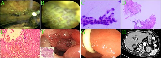
(A) Cholangioscopy revealed numerous common bile duct (CBD) stones. (B) A grid-like foreign body was identified within the CBD. (C) Brushing cytology was performed. (D) Cholangioscopy-guided biopsy pathology showed IgG (+), CD38 (+), and IgG4 (−) staining. (E) The pathology specimen from choledocholithotomy demonstrated chronic suppurative inflammation. (F) Colonoscopy revealed sigmoid colon cancer with a p53 missense mutation and a Ki-67 labeling index of 90%. (G) Two months post-surgery, endoscopy showed a significantly patent duodenal bulb. (H) CT indicated a more distinct interface between the hilar bile duct and liver segment 4 compared to prior imaging.
Subsequently, the patient underwent choledochotomy with T-tube drainage. Intraoperatively, multiple CBD stones and a hemostatic gauze-like foreign body were extracted, concomitant with a partial hepatectomy. Pathological examination of the resected hilar lesion revealed fibrotic nodules with calcification and chronic suppurative inflammation (Fig. 2E). One week later, colonoscopy confirmed sigmoid colon cancer (Fig. 2F). However, the patient and her family declined surgical resection or chemotherapy for the colon and renal masses, opting for supportive care only. Follow-up endoscopy two months postoperatively showed relief of the duodenal bulb stenosis (Fig. 2G), and CT demonstrated significant improvement in hilar bile duct inflammation (Fig. 2H). The patient died 8 months after the choledochotomy, as summarized in the timeline figure (Fig. 3).
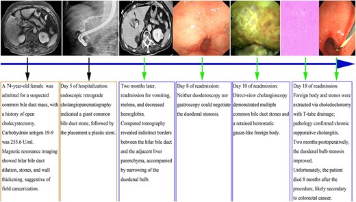
Timeline of diagnostic workup and surgical management. The figure outlines the clinical trajectory, including the initial endoscopic examination, the failure to traverse the duodenal stricture, and subsequent cholangioscopy revealing a retained hemostatic gauze. Following choledochotomy and foreign body extraction, the duodenal stenosis showed significant improvement; however, the patient passed away from non-biliary malignancies eight months post-choledochotomy.
Discussion
pCCA is a highly aggressive malignancy associated with a poor 5-year overall survival rate [6, 7]. Its diagnosis requires the integration of clinical, laboratory, imaging, and endoscopic evaluations [8]. The hallmark clinical presentation is obstructive jaundice, often accompanied by abdominal pain, pruritus, and weight loss [9]. Although laboratory findings typically reveal elevated levels of bilirubin, alkaline phosphatase, and CA19–9, these markers lack diagnostic specificity. For instance, elevated CA19–9 levels can be observed in benign hepatobiliary and pancreatic conditions, including primary sclerosing cholangitis (PSC), primary biliary cholangitis, chronic hepatitis, acute liver failure, cholangitis, and pancreatitis [10, 11]. This diagnostic dilemma is highlighted by the finding that at a CA19–9 cutoff of 32 U/ml, 54.9% of patients with benign jaundice tested positive [11]. While raising the CA19–9 cutoff threshold improves specificity, it inevitably compromises sensitivity [11]. In patients with PSC, a higher cutoff of 129 U/ml is required to achieve a specificity of 98.5% for diagnosing CCA, albeit with a reduced sensitivity of 78.6% [12]. Furthermore, CA19–9 is inherently undetectable in approximately 7% of the general population due to the absence of the Lewis antigen [9]. Given that IgG4-related sclerosing cholangitis closely mimics pCCA, assessing inflammatory markers such as IgG4 is also critical [13].
Imaging modalities—including ultrasound, contrast-enhanced CT/MRI, magnetic resonance cholangiopancreatography (MRCP), and positron emission tomography-CT—are fundamental to the diagnostic workup. However, imaging alone is insufficient, particularly considering that 3% to 15% of resected hilar strictures prove to be benign on final pathology [13, 14]. Consequently, endoscopic methods (such as endoscopic ultrasound-guided fine-needle aspiration [EUS-FNA], ERCP brush cytology, endobiliary biopsy, and cholangioscopy-guided biopsy) are employed to secure a tissue diagnosis. EUS-FNA demonstrates lower sensitivity for pCCA than for distal cholangiocarcinoma and is contraindicated in liver transplant candidates due to the risk of tumor seeding [9]. Regarding ERCP-based sampling, sensitivities remain limited: 45% for brush cytology, 48% for biopsy, and 59% for their combination [15]. Although cholangioscopy-guided biopsy improves sensitivity to 66% for indeterminate biliary strictures, it is considered a cost-effective strategy [15]. Other endoscopic modalities also present limitations: intraductal ultrasound lacks tissue acquisition capabilities, and probe-based confocal laser endomicroscopy is hampered by low specificity [15]. Ultimately, distinguishing pCCA from benign disease remains a persistent clinical challenge.
To our knowledge, this is the first reported case of chronic cholangitis mimicking pCCA secondary to retained calculi and a foreign body. We postulate that the foreign body was related to a previous cholecystectomy performed at a local hospital. Specifically, the conversion from laparoscopic to open cholecystectomy, likely necessitated by intraoperative hemorrhage, may have prompted the use of hemostatic gauze. The possible mechanism involved an inflammatory reaction caused by the retained gauze, followed by erosion through the wall of the extrahepatic bile duct.
In conclusion, differentiating pCCA from chronic inflammation remains challenging, particularly in the presence of surrounding organ involvement. Emerging technologies, such as fluorescent in situ hybridization, next-generation sequencing, and circulating cell-free DNA analysis, should be incorporated to enhance diagnostic accuracy.
Conflicts of interest
None declared.
Funding
None declared.


