


 https://orcid.org/0000-0003-0503-1773

Abstract
Pelvic lymphocele is a recognized complication of pelvic lymph node dissection (PLND), but fistulization to the sigmoid colon is an exceedingly rare sequela with limited management guidance. We report a 48-year-old woman who had undergone laparoscopic hysterectomy, bilateral salpingo-oophorectomy, and PLND for cervical cancer (pT1b3N1a, FIGO IIIC) and was found during adjuvant chemotherapy to have fever, left lower-quadrant pain, and computed tomography evidence of an infected pelvic lymphocele. Percutaneous drainage identified a 2-cm sigmoid colon fistula, but its size and anatomical risk precluded interventional radiologic closure. The patient underwent segmental sigmoid resection with end-to-side anastomosis and lymphocele marsupialization. Postoperative Enterococcus faecalis infection was managed with targeted antibiotic therapy, and follow-up imaging confirmed complete resolution. Early surgical intervention should be considered when conservative and interventional approaches are impractical in symptomatic pelvic lymphocele complicated by colonic fistulization.
Introduction
A lymphocele is a cystic lymphatic-fluid collection that may complicate pelvic lymph node dissection (PLND), with a reported incidence of 1%–58% after PLND for gynecologic malignancy [1]. Most lymphoceles are asymptomatic and resolve spontaneously, but symptoms develop in 5%–18% of cases [1–3]. Initial management of symptomatic lymphocele includes percutaneous fine-needle aspiration, catheter drainage, and embolization, while surgical fenestration or marsupialization is reserved for refractory cases, particularly after renal transplantation [3–6]. Progression to a fistula with the sigmoid colon is exceedingly rare, and cases requiring colectomy are rarer still. We report an infected pelvic lymphocele that fistulized to the sigmoid colon and required segmental sigmoid resection.
Case report
A 48-year-old woman with no significant medical history had undergone laparoscopic hysterectomy, bilateral salpingo-oophorectomy, and PLND for cervical cancer (pT1b3N1a, FIGO IIIC). She received four cycles of paclitaxel and cisplatin, with a course complicated only by lower-extremity edema. Surveillance abdominal computed tomography (CT) before the fifth cycle showed a pelvic lymphocele, which was managed conservatively under serial observation (Fig. 1).
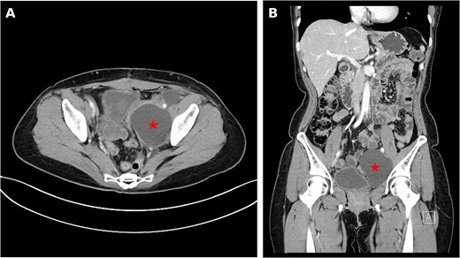
Initial detection of pelvic lymphocele on surveillance imaging. (A) Axial contrast-enhanced abdominopelvic CT image and (B) coronal reconstruction obtained before the fifth cycle of adjuvant chemotherapy, demonstrating a left pelvic lymphocele (red star) without rim enhancement or surrounding inflammatory change.
On scheduled admission for the fifth cycle, the patient presented with a temperature of 38.5°C and left lower-quadrant pain. White blood cell count was 9330/μL (neutrophils 79.6%) and C-reactive protein 12.47 mg/dL. Chemotherapy was withheld, and intravenous cefepime 2 g every 8 h was initiated. Repeat CT showed enlargement of the lymphocele with intralesional gas tracking from the adjacent sigmoid colon, raising suspicion for infection and colonic fistulization (Fig. 2). Sigmoidoscopy was attempted but could not be completed due to patient discomfort and did not identify a fistula. Percutaneous drainage was performed for both diagnosis and decompression, and contrast study revealed a 2-cm-wide fistulous tract between the lymphocele and the sigmoid colon (Fig. 3). Coil embolization and glue injection were considered but deemed infeasible because of fistula size and the risk of intracolonic spillage of sclerosing agent. The colorectal surgery service was consulted.
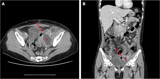
Follow-up CT at the time of clinical deterioration. (A) Axial contrast-enhanced CT image and (B) coronal reconstruction showing interval enlargement of the lymphocele (red star) with intralesional gas (red arrow) tracking from the adjacent sigmoid colon, suggesting infection and fistulization.
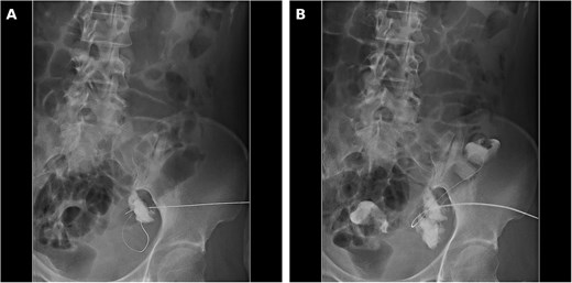
Fluoroscopic images obtained during percutaneous catheter drainage of the lymphocele. (A) Initial contrast injection opacifies the lymphocele cavity. (B) Continued contrast injection demonstrates a 2-cm-wide fistulous tract with opacification of the adjacent sigmoid colon lumen.
The patient underwent segmental sigmoid resection with end-to-side anastomosis and lymphocele marsupialization. Intraoperatively, dense adhesions were noted between the pelvic cavity, the sigmoid colon, and the lymphocele. The involved segment was meticulously dissected free, resected with an 80-mm linear stapler, and an end-to-side anastomosis was created with a 28-mm circular stapler. The lymphocele cavity was widely opened and curetted, and Jackson-Pratt (JP) drains were placed in the pelvic cavity and the lymphocele bed.
Postoperatively, the patient resumed sips of water at 6 h and a soft diet on Day 4 after passing flatus. Persistent fever ≥38.5°C despite cefepime prompted culture of the drainage fluid, which grew Enterococcus faecalis. Antibiotics were narrowed to ampicillin on postoperative Day 3 per infectious-disease consultation, and the fever defervesced by Day 5. The pelvic JP drain was removed once output decreased and became serous; the lymphocele-bed JP drain was maintained, and the patient was discharged on suppressive drainage. On postoperative Day 24, the patient was readmitted for the sixth chemotherapy cycle, the remaining drain was removed, and follow-up CT confirmed complete resolution of the lymphocele without anastomotic complication (Fig. 4).
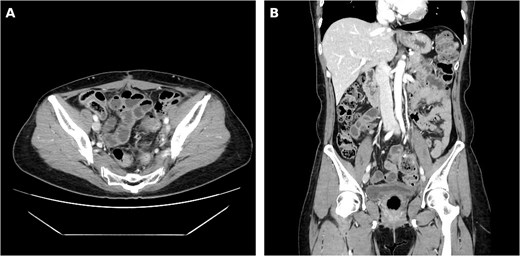
Postoperative follow-up imaging obtained 24 days after surgery. (A) Axial contrast-enhanced CT image and (B) coronal reconstruction demonstrating complete resolution of the lymphocele and a patent sigmoid colon at the anastomotic site without leak or residual collection.
Discussion
Pelvic lymphocele is an abnormal lymphatic-fluid collection most commonly seen after pelvic lymphadenectomy for gynecologic, urologic, or renal malignancy, or after renal transplantation [7]. Reported risk factors include high body mass index, omission of lymphatic ligation, extensive lymphadenectomy, anticoagulant use, and prior radiotherapy [8–11]. Most lymphoceles remain asymptomatic and resolve spontaneously, but 5%–10% become symptomatic from mass effect on adjacent rectum, bladder, ureter, or vessels, manifesting as pain, urinary symptoms, edema, or deep vein thrombosis [13, 14]. Lymphoceles typically develop 3–8 weeks postoperatively but may appear up to one year later and mimic recurrence [9, 12]. CT is preferred for diagnosis and surveillance, allowing assessment of complications and image-guided intervention [5, 15]. The differential diagnosis includes hematoma, abscess, urinoma, seroma, and cystic neoplasm.
When complications arise, conservative management or interventional radiology remains first-line, and surgical intervention is reserved for refractory cases. Reports of lymphocele-to-sigmoid fistulization are exceedingly scarce, and to our knowledge optimal management is undefined. In our case, the fistula was too wide for occlusion by drainage alone, and sclerosing agents (ethanol, bleomycin) posed a risk of intracolonic spillage. Endoscopic stenting was considered but abandoned because of anatomical migration risk. Segmental sigmoid resection was therefore unavoidable.
In retrospect, the only sentinel feature at the time of initial lymphocele detection was lower-extremity edema, and the gynecology team appropriately elected watchful waiting given the natural history of spontaneous resolution. This case nonetheless illustrates that, once a lymphocele is identified, close surveillance with serial imaging and prompt multidisciplinary consultation upon any symptom progression are essential, with a low threshold for interventional or surgical treatment when less-invasive approaches are not feasible.
Conflicts of interest
The authors declare that they have no conflicts of interest.
Funding
This research received no specific grant from any funding agency in the public, commercial, or not-for-profit sectors.
Data availability
No datasets were generated or analyzed during the current study. All relevant information is included in this article.
Ethics approval
This study was approved by the Institutional Review Board of Inha University Hospital with a waiver of informed consent (IRB No. 2026-04-006). Written informed consent was obtained from the patient for the publication of this case report and any accompanying images.


