


 https://orcid.org/0000-0002-6904-7267

Abstract
Middle mediastinal schwannomas are rare and can mimic malignant mediastinal tumours, making preoperative diagnosis challenging. A 52-year-old woman presented with persistent cough refractory to medical treatment. Chest computed tomography revealed a well-defined 35 × 26 mm mass in the right middle mediastinum, and 18F-fluorodeoxyglucose positron emission tomography showed increased uptake with a maximum standardized uptake value of 8.8. Endobronchial ultrasound-guided transbronchial needle aspiration was performed, and histological examination revealed S-100-positive spindle-shaped cells with a low Ki-67 labeling index, leading to a diagnosis of schwannoma. Based on the tumour location and pathological diagnosis, the cough was considered attributable to vagal nerve irritation. Thoracoscopic tumour resection was performed with careful preservation of the vagal nerve. The cough resolved promptly after surgery, and no recurrence was observed during 1 year of follow-up. Preoperative pathological diagnosis was useful for planning a nerve-preserving surgical strategy.
Introduction
Schwannomas are benign tumours originating from Schwann cells and are among the most common neurogenic tumours of the mediastinum. However, they usually arise in the posterior mediastinum, whereas middle mediastinal schwannomas are rare. Because middle mediastinal tumours may mimic malignant diseases such as thymoma, thymic carcinoma, and malignant lymphoma, diagnosis based solely on imaging findings is often challenging [1, 2]. Endobronchial ultrasound-guided transbronchial needle aspiration (EBUS-TBNA) has recently become a minimally invasive diagnostic modality for mediastinal lesions [3].
Case presentation
The patient was a 52-year-old woman, 156 cm tall, weighing 77.0 kg, with a body mass index of 31.6 kg/m2, who presented with a persistent cough. Her medical history included childhood bronchial asthma and gastroesophageal reflux disease. There was no significant family history. She had a smoking history of 10 cigarettes per day between the ages of 20 and 23 years.
Two weeks before presentation, she noticed continuous cough and visited a local clinic. Because she had a history of childhood bronchial asthma, an asthma exacerbation was suspected, and medical treatment was initiated; however, her symptoms did not improve. Chest imaging performed for further evaluation revealed a middle mediastinal mass, and she was referred to our department for surgical management. She was alert and haemodynamically stable. Breath sounds were clear bilaterally, and heart sounds were normal. No cervical or supraclavicular lymphadenopathy was detected. Laboratory examinations, including hematological, biochemical, and coagulation tests, showed no abnormalities. Tumour markers were within normal limits.
Chest radiography showed no infiltrative shadows or nodular lesions in the lung fields. Chest computed tomography (CT) revealed a well-defined, homogeneous mass measuring 35 × 26 mm in the right middle mediastinum, without evidence of invasion into surrounding structures (Fig. 1). Chest magnetic resonance imaging was not performed at the patient’s request. 18F-Fluorodeoxyglucose-positron emission tomography revealed high accumulation in the same area, with a maximum standardized uptake value of 8.8 (Fig. 2). No significant uptake or enlargement was observed in mediastinal or hilar lymph nodes. Based on these findings, thymoma, thymic carcinoma, and malignant lymphoma were considered in the differential diagnosis.
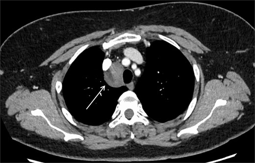
Chest CT findings. A well-defined, homogeneous mass measuring 35 × 26 mm was observed in the right middle mediastinum (arrow), with no evidence of invasion into the surrounding structures.
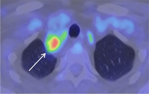
18F-fluorodeoxyglucose-positron emission tomography findings. Increased FDG uptake was observed in the middle mediastinal mass, with a maximum standardized uptake value (SUV max) of 8.8.
For definitive diagnosis, EBUS-TBNA was performed. Histopathological examination revealed proliferation of spindle-shaped cells, and immunohistochemical staining showed positivity for S-100 protein with a low Ki-67 labeling index, leading to a diagnosis of schwannoma (Fig. 3A–D). Biopsy results indicated the cough was due to tumour-related nerve compression, so surgery was scheduled. Prior to surgery, the necessity of nerve preservation was carefully discussed within the surgical team.
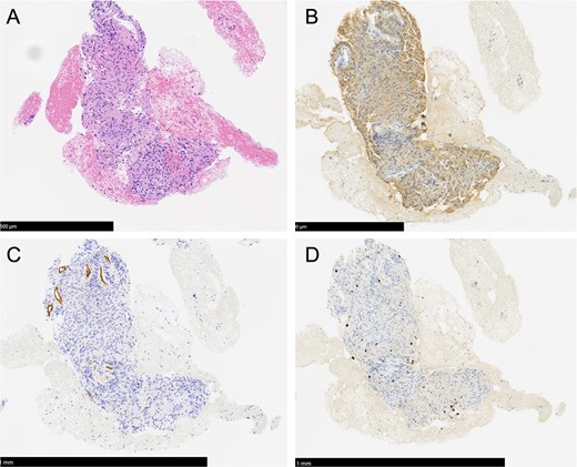
Histopathological findings of endobronchial ultrasound–guided transbronchial needle aspiration (EBUS-TBNA). (A) Hematoxylin–eosin (HE) staining demonstrates proliferation of spindle-shaped cells arranged in fascicles, consistent with an Antoni A pattern (scale bar = 500 μm). (B) Immunohistochemical staining shows diffuse positivity for S-100 protein in tumor cells (scale bar = 500 μm). (C) Tumor cells are negative for CD34 (scale bar = 1 mm). D. The Ki-67 (MIB-1) labeling index is low, indicating low proliferative activity (scale bar = 1 mm).
Thoracoscopic surgery was performed under general anaesthesia with the patient in the left lateral decubitus position. No intraoperative nerve monitoring system was used. Because schwannoma had been diagnosed preoperatively by EBUS-TBNA, a nerve-preserving surgical strategy was planned before surgery. The tumour was located adjacent to the right vagus nerve, and nerve-preserving tumour resection was performed with meticulous identification of the nerve course to preserve nerve function (Fig. 4A and B). Under thoracoscopic visualization, the vagus nerve was carefully identified, and the tumour was dissected along the tumour capsule while preserving the macroscopic continuity of the nerve. The recurrent laryngeal nerve was not directly exposed in the operative field. To minimize thermal injury, electrocautery and energy devices were avoided as much as possible around the vagus nerve. Dissection near the nerve was performed mainly using blunt and sharp dissection, and hemostasis was achieved primarily by compression and clipping. The surgery lasted 88 min, and the amount of bleeding was minimal. Postoperatively, the patient showed no hoarseness, dysphagia, aspiration, or other symptoms suggestive of recurrent laryngeal nerve palsy.
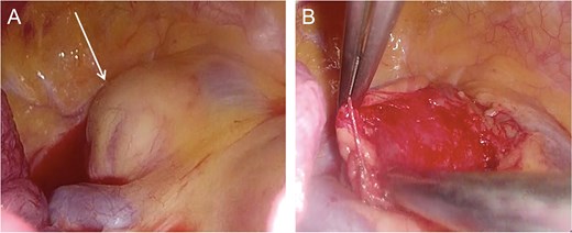
Intraoperative findings. Thoracoscopic intraoperative views. (A) The tumour was identified in the middle mediastinum with a smooth surface. (B) The tumour was adjacent to the vagal nerve. Nerve-preserving tumour resection was performed following meticulous identification of the nerve course.
Histopathological examination of the resected specimen revealed bundles of spindle-shaped cells predominantly exhibiting an Antoni A pattern. Based on the histological and immunohistochemical findings, the final diagnosis of schwannoma was confirmed (Fig. 5A and B). The postoperative course was uneventful, and the patient was discharged on postoperative day 7. The persistent cough resolved promptly after surgery, and no recurrence has been observed during 1 year of follow-up.
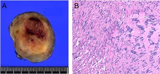
Histopathological findings of the resected specimen. (A) Gross findings of the resected tumour. (B) HE staining shows interlacing spindle-shaped cells with alternating hypercellular and hypocellular areas, consistent with Antoni A and Antoni B patterns (×10).
Discussion
Middle mediastinal schwannomas are rare and may be difficult to diagnose preoperatively because they can mimic other mediastinal lesions. In the evaluation of middle mediastinal masses, lymphadenopathy, lymphoid malignancies, metastatic lymph nodes, bronchogenic or duplication cysts, and vascular lesions are usually considered in the differential diagnosis. Although neurogenic tumours typically arise in the posterior mediastinum, they should also be considered when a middle mediastinal mass is located along the course of the vagus nerve [4]. Recent reviews on mediastinal lesions have emphasized that imaging findings alone are often insufficient to determine benignity/malignancy or histologic subtype, highlighting the need for structured, multimodal assessment [5–7]. Therefore, precise lesion localization and clinical features, including symptom patterns, should be integrated to include neurogenic tumours in the differential diagnosis.
Schwannomas arising from the vagal or recurrent laryngeal nerve may cause neurogenic irritation symptoms, including cough, hoarseness, and dysphagia, depending on tumour location and growth direction. Recent case reports have described mediastinal schwannomas detected after respiratory or chest symptoms, including persistent cough; although nonspecific, these symptoms may provide diagnostic clues when correlated with tumour anatomy [8, 9]. In the present case, persistent cough resolved promptly after resection, suggesting tumour-related vagal nerve irritation. Therefore, cough associated with an middle mediastinal mass should not be regarded solely as an incidental respiratory symptom, but may also prompt consideration of a neurogenic tumour.
In the present case, EBUS-TBNA enabled preoperative diagnosis of schwannoma. Given that middle mediastinal schwannomas often cannot be distinguished from malignant tumours on imaging alone, pathological confirmation prior to treatment is valuable for determining an appropriate management strategy. Previous reports have demonstrated the utility of EBUS-TBNA for diagnosing mediastinal neurogenic tumours, and the current case extends its application to middle mediastinal lesions [10, 11]. However, EBUS-TBNA may be limited by the amount of tissue obtained, particularly in benign mediastinal conditions. Recently, endobronchial ultrasound–guided intranodal forceps biopsy using standard-sized biopsy forceps has been shown to improve diagnostic yield for mediastinal lesions, including benign disease, and may become a useful diagnostic modality for middle mediastinal schwannomas in the future [12, 13].
Surgical resection remains the cornerstone of treatment for schwannomas. However, tumours arising from the vagal or recurrent laryngeal nerve present distinct challenges due to the risk of postoperative nerve dysfunction [14]. Although nerve-preserving approaches have been reported, transient or permanent recurrent laryngeal nerve palsy may occur [15]. In the current case, resolution of persistent cough after surgery highlights the clinical significance of resection. In benign tumours, a strategy of nerve-preserving tumour resection, rather than complete nerve sacrifice, is appropriate to optimize postoperative quality of life. Preoperative pathologic confirmation allowed the selection of a minimally invasive thoracoscopic approach, avoiding more extensive surgery.
In conclusion, neurological symptoms such as persistent cough should prompt consideration of neurogenic tumours in the differential diagnosis of middle mediastinal masses. Preoperative pathological diagnosis using EBUS-TBNA is a valuable, minimally invasive approach that facilitates appropriate surgical planning, including nerve-preserving procedures.
Author contributions
Nozomi Hatanaka (Conceptualization, Validation, Writing—original draft, Writing—review & editing), Ryusei Yoshino (Conceptualization, Validation, Visualization, Writing—original draft, Writing—review & editing), Miku Tamiya (Validation), Takumi Inao (Validation), Kengo Takahashi (Validation), Akane Ito (Validation), Shunsuke Yasuda (Validation), and Masahiro Kitada (Validation, Supervision). All authors have read and approved the final version of the manuscript.
Conflicts of interest
All authors have no competing interests, financial or non-financial.
Funding
This study received no funding from the public, private, or commercial sectors.
Data availability
Data sharing is not applicable to this article because no datasets were generated or analyzed in the current study.
Ethics approval
The study was conducted in accordance with the guidelines of the Declaration of Helsinki and was approved by the Institutional Review Board of Asahikawa Medical University Hospital (No. 25108). Approval date: March 8, 2026.
Consent for publication
Written informed consent was obtained from the patient for the publication of this study.


