


Abstract
Transoral thyroidectomy via vestibular approach has gained increasing acceptance due to its excellent cosmetic outcomes. Transoral robotic thyroidectomy (TORT) represents a further technical evolution, potentially offering enhanced options of instrument movement and visualization. However, no reports from Europe were yet published. We report the first TORT performed in Germany in a 56-year-old female presenting with a symptomatic 20 × 16 mm sized nodule in the right thyroid lobe measuring 14 ml. A right hemithyroidectomy was performed using TORT with the daVinci Xi® robotic system. Total operative time was 230 min. The postoperative course was uneventful, and the patient was discharged on postoperative day 3. TORT is feasible and safe in selected patients also in the German hospital setting, although the additional costs are not reimbursed by the German health system. This case demonstrates the technical applicability and highlights key procedural steps for implementation.
Introduction
Transoral thyroidectomy via the vestibular approach (TOETVA) is a technique that avoids visible cervical scarring by utilizing a natural orifice access [1, 2]. Since its introduction, it has gained worldwide acceptance and has also been implemented in very few centers in Germany during the last 8 years [3–6]. Transoral robotic thyroidectomy (TORT) represents a further evolution of this approach, combining the advantages of TOETVA with enhanced visualization and articulating instruments provided by a robotic system. While conduction of TORT in humans has been reported mainly from highly specialized Asian centers [7–11], there are yet no published reports from Europe. To the best of our knowledge, we present the first TORT performed in Germany.
Case report
A 56-year-old, healthy female patient presented with a palpable thyroid nodule in the right lobe which caused swallowing discomfort. Neck ultrasound revealed a 20 × 16 mm sized nodule, classified as EU-TIRADS III, in the upper pole of the right thyroid lobe with a total volume of 14 ml. The patient decided against a proposed fine-needle aspiration cytology of the nodule. She wanted resection of the symptomatic nodule, and no radiofrequency ablation. The preoperative vocal cord examination revealed no abnormalities. The patient was counseled regarding the surgical options for right hemithyroidectomy, and she opted for TORT after informed consent.
Surgical technique
The operation set up is shown in Fig. 1. The patient was positioned in a beach-chair position and orally intubated with an electromyogram endotracheal tube (Dr. Langer Medical). A single-shot antibiotic prophylaxis with ampicillin/sulbactam was administered. After general anesthesia was induced, the head was placed on a headplate without head fixation, eye protection flaps were placed, and the head was wrapped with a soft towel, which was overtaped with surgical incision film (Fig. 2A). Subsequently, her oral cavity was irrigated with a solution of octenisept® antiseptic spray. Skin disinfection and draping was performed to expose an operation field from the lower lip to the sternal notch (Fig. 2B).
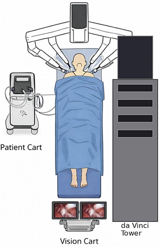
Operation set-up.
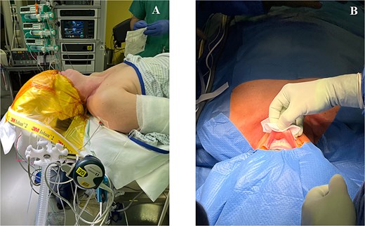
Positioning and preparation of the patient. (A) Head placed on a headplate without head fixation, head overtaped with surgical incision film and (B) Disinfected and draped operation field from the lower lip to the sternal notch.
Access and working space creation
A median incision of 10 mm was made in the mucosa of the lower lip very close to the lip vermilion (Fig. 3A). Through the median incision, the musculature was bluntly separated using a clamp and dissected toward the inferior border of the mandible, thereby entering the subplatysmal space. Using a bluntly advanced Veress needle, about 300 ml hundred milliliters of a prepared solution (500 ml normal saline with 1–2 mg epinephrine) was injected to create a relatively bloodless working space between the platysma and the strap muscles. Prior to trocar insertion, this space was further developed using a blunt dissector or tunneling instrument in a fan-shaped manner in three directions: medially in the pretracheal plane and laterally to both sides, thereby creating three tunnels that could be easily connected. Subsequently, the medial 8-mm da Vinci trocar was inserted (Fig. 3B). Thereafter two lateral 5–8 mm incisions in the vestibulum of the lower lip were made as lateral as possible to create enough space for the two lateral 8 mm daVinci trocars. A sufficient distance between trocars seems to be essential to ensure trouble-free use of the robotic arms without collisions. Again, a bluntly advanced Veress needle with the prepared saline-epinephrine solution was used for hydrodissection before insertion of the two lateral trocars. Then CO₂ insufflation was initiated (maximum pressure 6–8 mmHg). The ventral placement of the trocars within the oral vestibule helps to preserve the mental nerve and the lower row of teeth.
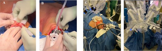
Access, trocar placement, and docking. (A) Median incision of 10 mm was made in the mucosa close to the lower lip; (B) insertion of the medial 8-mm da Vinci trocar and lateral incision; (C) insertion of the two lateral trocars and further creation of the subplatysmal working space; and (D) docking of the robotic arms over the top.
Following trocar placement, the previously created subplatysmal working space was further developed with conventional laparoscopic instruments by connecting the three trocar tunnels under direct visualization (Fig. 3C) until the strap muscles are fully exposed, extending from the sternal notch to the thyroid cartilage and laterally to the sternocleidomastoid muscles. After creation of the subplatysmal space the daVinci XI (Intuitive Surgical®), patient-side cart was docked (Fig. 3D).
Console phase
Dissection of the thyroid gland was initiated by longitudinal division of the strap muscles with the harmonic scalpel (Harmonic ACE®), allowing visualization of the trachea and thyroid isthmus (Fig. 4A). The right strap muscles are then retracted laterally using one percutaneous stay suture to optimize the operative field. The carotid sheath, containing the common carotid artery, internal jugular vein, and vagus nerve were identified by blunt dissection. This allowed intermittent intraoperative neuromonitoring (IONM) of the vagus nerve prior to resection.
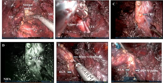
Console phase. (A) View of the subplatysmal operation field; (B) mobilization of the upper pole and clipping of the superior thyroid artery (STA); (C) mobilized upper thyroid pole with upper parathyroid gland (UPG); (D) near-infrared fluorescence angiography of the UPG; (E) visualization RLN at the entry point of the RLN into the laryngeal musculature; (F) IONM of the RLN with a long stimulation probe. SM- sternocleidomastoid muscle.
Thyroid dissection began with division of the isthmus, followed by mobilization of the superior pole. After opening the avascular plane between the superior pole and the laryngeal musculature, the superior thyroid artery was dissected free, clipped with a resorbable clip (Fig. 4B) and divided using an energy device. The superior pole was then mobilized ventro-medially to expose the superior parathyroid gland (Fig. 4C and D) and the entry point of the recurrent laryngeal nerve (RLN) into the laryngeal musculature (Fig. 4E), which was confirmed by intraoperative neuromonitoring with a long probe (Fig. 4F). The RLN was carefully dissected from the posterior surface of the thyroid gland and the lateral attachments. Then, two terminal branches of the inferior thyroid artery, and the middle thyroid vein (Kocher vein) were divided under direct vision with the Harmonic ACE (Intuitive Surgical®) or with a scissors after clip placement. After division of Berry’s ligament, the thyroid lobe became fully mobile and could be retracted toward the contralateral side. The inferior parathyroid gland was then identified and dissected laterally preserving its blood supply.
Once the specimen was completely mobilized, it was retrieved via the incision of the central trocar using an endoscopic retrieval bag. The weight of the specimen was 15 g.
After specimen retrieval hemostasis was assessed by increasing intrathoracic pressure up to 40 mmHG via ventilation. The intact function of the vagus nerve and RLN was confirmed to be intact by IONM at the end of the procedure. The operating field was finally rinsed with normal saline. The strap muscles were not reapproximated. The mucosal incisions were closed meticulously with rapidly absorbable 5.0 sutures. The total operating time was 230 min.
The final pathological report stated a 16 × 21 mm sized follicular adenoma without signs of malignancy.
Postoperative course
The postoperative course was uneventful, despite a slight hypoesthesia around the chin, which resolved after 14 days. Serum calcium and parathyroid hormone levels at postoperative day 1 were within normal limits. The patient was discharged at postoperative day 3. The laryngoscopic assessment of the vocal cord function with a stroboscope by an ENT specialist at postoperative day 7 showed a slightly reduced mobility of the right vocal cord, which has been resolved after logopedic treatment at the 3 months control. At the outpatient control examination at postoperative day 14, the patient was well satisfied with the cosmetic result (Fig. 5A and B).
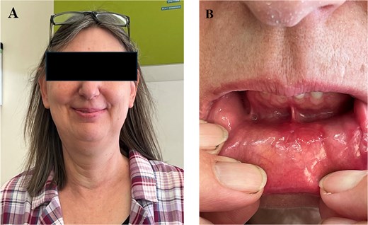
Postoperative cosmetic result at day 14: (A) neck; (B) mouth vestibulum.
Discussion
To best of our knowledge, we report the first TORT performed in Germany. Although TORT became a well-established procedure in some specialized centers in Asia [6–9], it has yet not been reported from Germany. There are several potential reasons for this fact. First, most endocrine surgeons in Germany either do not have access, nor are familiar with the use of a robotic system. Secondly, the operating time is approximately 3 times longer than in conventional open surgery, which is a major economic point given the lack of operation capacity in the majority of surgical departments. Third, the TOETVA/TORT procedure is more expensive than conventional thyroidectomy, but not refinanced by the German health system.
As TOETVA also TORT provides an excellent cosmetic outcome, as in the present case, by utilizing natural orifice for its access. Intraoral scars are not only hidden from the exterior, but also completely disappear in a few months [1–9]. TORT carries several potential advantages in technical aspects compared to TOETVA, including enhanced 3D visualization, improved dexterity with articulating instruments and better ergonomics [6–9]. Therefore, it was decided to use the TORT approach. Robotic EndoWrist technology also allows TORT to have deeper approach into the superior pole for complete dissection. In addition, TORT has flexible counter-traction around peri-neural tissue near the RLN and the trachea which allows complete dissection of ligament of Berry. Both postulated advantages could be experienced in the presented case. During preparation of the thyroid lobe also the SynchroSeal sealing device (Intuitive Surgical®) was used, because of its better movement degrees. However, compared to the Harmonic ACE, the instrument was too big and the branches to crude in the narrow operating field, so that it was more comfortable to proceed with the harmonic scalpel. TORT is also associated with an increased complexity compared to TOETVA. The console surgeon has carefully to perform small movements to avoid tearing the lower lip and injuring the lower teeth. The range of robotic arm movement must be careful monitored by the table assistant all the time, since this seems to be crucial.
The operative time in the presented case was in the range of the literature, keeping in mind that operative times for TORT are reported to be about 15 min longer than for TOETVA [10]. The presented patient experienced a transient palsy of the recurrent laryngeal nerve, which occurs according to recent meta-analysis in 0%–2.48% of TORT compared to 1.3%–9.3% of TOETVA procedures [11]. Thus, transient recurrent laryngeal nerve palsy was lower in the TORT group than in the TOETVA group (OR 2.69; 95% CI 1.09–6.62), but other complications such as bleeding, infection, hypoparathyroidism, and injury of the mental nerve were not different.
Conclusion
Robotic transoral thyroid resection is feasible in a German clinical setting and thus represents one further alternative for thyroid resection.
Conflicts of interest
None declared.
Funding
None declared.
Consent
Written informed consent was obtained from the patient for publication of this case report and her photographs.


