


 https://orcid.org/0000-0003-3624-6005

Abstract
Perivascular epithelioid cell tumors (PEComas) are rare mesenchymal neoplasms composed of distinctive perivascular epithelioid cells that exhibit both melanocytic and smooth muscle differentiation on immunohistochemistry. These tumors can develop in various anatomic locations and display variable biological behavior ranging from benign to aggressive malignant disease. We present a case series of four patients diagnosed with PEComa at different anatomic sites. Histopathological analysis in all cases demonstrated characteristic epithelioid or spindle-shaped morphology, with some cases showing positive staining for melanocytic markers such as HMB-45 and Melan-A, as well as smooth muscle markers. Due to their rarity, definitive management protocols are not yet well established. Surgical resection remains the primary treatment for localized disease, while systemic therapies targeting the mammalian target of rapamycin pathway may be considered for unresectable tumors or metastatic disease. This case series underscores the heterogeneity of PEComas and highlights the importance of multidisciplinary management and long-term follow-up.
Introduction
Perivascular epithelioid cell tumors (PEComas) belong to a rare group of mesenchymal neoplasms composed of distinctive PECs that exhibit unique immunoreactivity for melanocytic and smooth muscle markers [1]. Histologically, PEComas have a perivascular distribution and are characterized by epithelioid to spindle cells with eosinophilic to clear cytoplasm and demonstrate positive immunostaining for melanocytic markers such as HMB45 and Melan-A, as well as for smooth muscle markers including smooth muscle actin, desmin, and SMA [1, 2].
The PEComa family includes angiomyolipoma (AML), lymphangioleiomyomatosis, and clear cell ‘sugar’ tumors of the lung (CCST), in addition to PEComa-not otherwise specified (PEComa-NOS) found in soft tissues and visceral organs [2, 3]. These tumors may develop in a broad range of anatomical locations, including the uterus, retroperitoneum, liver, gastrointestinal tract, pancreas, and soft tissues of the extremities [4]. Most cases occur between the ages of forty and sixty, with a higher prevalence in females, resulting in an overall female-to-male ratio of 4: 1 [1, 5, 6].
Due to their rarity, the natural history of PEComas and optimal management strategies remain uncertain, and it has not yet been possible to fully establish criteria for malignancy [6]. Surgical resection is generally considered the primary treatment for localized disease, whereas systemic therapies, particularly inhibitors of the mammalian target of rapamycin (mTOR) pathway, have shown promising activity in metastatic or unresectable cases [5–9].
In this report, we present a case series of four patients with PEComa involving the lower extremity, uterus, and liver, illustrating the diverse clinical presentations and management strategies of this rare tumor.
Case series
Case 1
A 70-year-old female presented with a painless, enlarging mass of ~4 × 7 cm along the anterior aspect of the right lower extremity for 6 months. Biopsy suggested the lesion may represent a PEComa. The patient underwent a wide local excision. Histopathologic evaluation was consistent with PEComa, showing a proliferation of nested epithelioid cells with clear cytoplasm that exhibited no significant cytologic atypia, mitoses, or necrosis (Fig. 1a and b). The cells were weakly positive for desmin and negative for HMB-45, Melan A, and SMA. Margins were clear. Given the benign nature of the PEComa and the R0 resection, the patient did not require any further treatment. She remains without evidence of recurrence after three years.
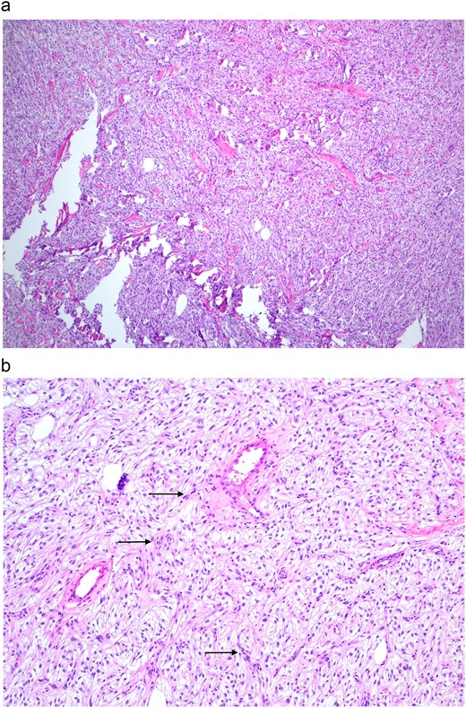
(a) H&E stain 40×: Dermal soft-tissue PEComa with eosinophilic and granular spindle-cell morphology. (b) H&E stain 100×: Elongated spindle-shaped cells with clear cytoplasm in fascicular arrangements (arrows), exemplifying the multiple morphologies that neoplastic cells can display.
Case 2
A 43-year-old female was incidentally found to have a 5-cm left-lobe liver mass with normal CA 19–9, CEA, and AFP levels (Fig. 2a and b). Percutaneous biopsy confirmed a PEComa expressing characteristic immunohistochemical markers, including HMB45 and SMA. The patient had numerous severe underlying medical comorbidities. Upon discussion at our institution’s multidisciplinary cancer conference, it was felt that, while surgical resection would be optimal, the potential morbidity associated with a major hepatic resection needed to be considered in this case. Due to the absence of symptoms and the lack of aggressive histologic features, it was elected to proceed with close watchful waiting with biannual serial imaging surveillance. Follow-up imaging at three years since diagnosis demonstrated stable disease without progression.
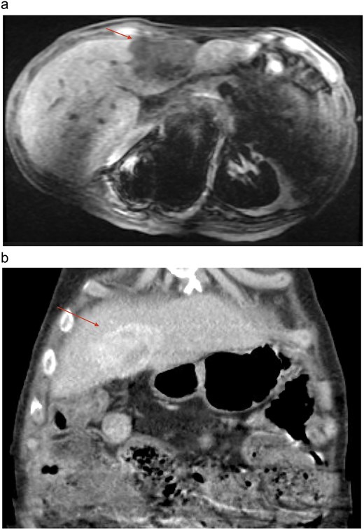
(a) MRI, axial section showing a heterogeneously enhancing subcapsular mass in the left hepatic lobe measuring 5.3 cm (arrow). (b) CT scan coronal section demonstrating a hypervascular lesion in the medial segment of the left liver lobe (arrow).
Case 3
A 38-year-old female with a history of fibromas presented with severe abdominal pain. A computed tomography (CT) scan demonstrated a large, 12.4 cm mass originating from the upper part of the uterine fundus, along with infiltration of the mesentery within the pelvis (Fig. 3). A Magnetic resonance imaging (MRI) 6 months after the initial CT scan showed that the mass had increased to 16.5 cm and contained internal septations. The patient underwent a total abdominal hysterectomy with bilateral salpingectomy. Final pathology identified a uterine PEComa characterized by cells with predominantly spindle cell morphology and a minor mixed component with epithelioid differentiation, exhibiting moderate to marked atypia, increased mitotic activity (up to 5 per 10 high-power fields), and necrosis. No lymph vascular invasion or infiltrative pattern were visualized. Immunohistochemical analysis showed the neoplastic cells were positive for HMB-45, Melan-A, actin, desmin, and cathepsin K, with increased Ki-67 and p53 expression (Fig. 4a and b). Based on the combined features, this PEComa was deemed to have uncertain malignant potential, and close surveillance was recommended, with imaging initially every six months and then annually. The patient remains disease-free after four years of surveillance.
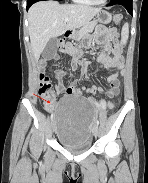
CT scan, coronal section demonstrating a large 12.4 cm mass arising from the superior aspect of the uterine fundus with infiltration of the mesentery within the pelvis (arrow).
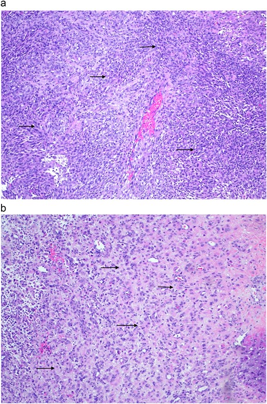
(a) H&E stain 100×: Elongated crowded spindle-shaped cells (arrows). (b) H&E stain 100×: Epithelioid cells exhibiting clear to eosinophilic and granular cytoplasm (arrows).
Case 4
A 19-year-old male presented with a painless, rapidly enlarging, firm, mobile, non-compressible soft-tissue mass ~4 cm in the right anterior lower extremity, with overlying skin darkening for one year. The patient underwent a wide local excision. Histology revealed epithelioid and spindle cells growing in sheets and nests around prominently hyalinized vasculature, with no evidence of mitotic activity or necrosis (Fig. 5a–c). Surgical margins were clear. Immunohistochemical staining showed positivity for cathepsin-K, SMA, and desmin. The morphological characteristics, along with the immunophenotype, supported the diagnosis of PEComa. In the absence of any aggressive features and following R0 resection, no additional treatment was deemed necessary. The patient has a follow-up appointment scheduled for 6 months post-surgery.
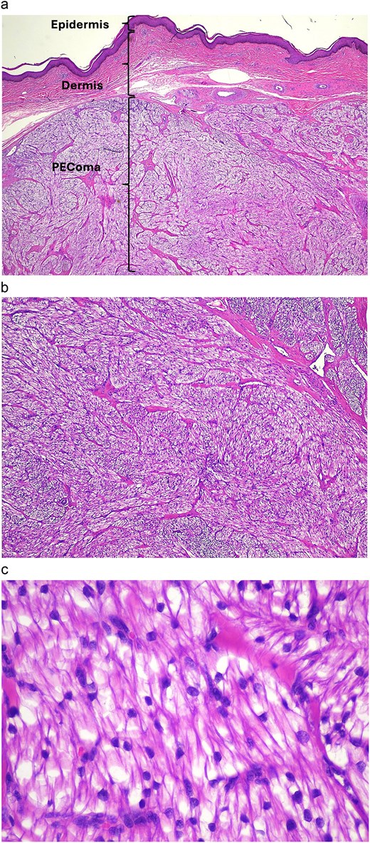
(a) H&E stain 20×: PEComa located within the dermis. The epidermis lines the superficial surface, while the uninvolved dermis lies beneath the epidermis. (b) H&E stain 40×: PEComa cells with clear to eosinophilic cytoplasm arranged in sheets and nests around the vasculature. Minimal mitotic activity noted. (c) H&E stain 400×: tumor cells showing clear to lightly eosinophilic cytoplasm, fine chromatin, and inconspicuous nucleoli.
Discussion
In 2002, the World Health Organization defined PEComa as ‘a mesenchymal tumor composed of histologically and immunohistochemically distinctive perivascular epithelioid cells’ [10]. However, the term PEC was initially introduced by Bonetti et al. [11], a decade earlier, in 1992, after noticing that both AML and CCST were composed of this unconventional cell type. This cell type is epithelioid in morphology, with clear to eosinophilic cytoplasm, a perivascular arrangement, and expression of melanocytic and muscle markers. Although no normal histologic counterpart has currently been recognized for the PEC [3], the unique phenotypic biology of PEComas has led to its identification in soft tissue, viscera, and several other organ sites [4]. These neoplasms should be meticulously differentiated from their morphological and immunohistochemical mimics, especially melanoma/clear cell sarcoma, gastrointestinal stromal tumors, and true smooth muscle tumors [6]. Since their initial recognition as a distinct pathological entity, PEComas have been increasingly documented across various organ systems. Nevertheless, their clinical behavior and optimal management remain incompletely understood owing to their rarity.
There is a strong association between PEComas and mutations in the tuberous sclerosis complex (TSC1) or (TSC2) genes, which are critical components of the mTOR signaling pathway. Of note, the connection between TSC and the rarer PEComas-NOS is less clear [10]. Around 20% of PEComas do not exhibit TSC1/TSC2 mutations and are instead characterized by translocations involving the TFE3 gene, which are usually associated with a more aggressive clinical behavior [8, 12].
The clinical presentation is often nonspecific and varies according to the tumor’s location and size [13]. Soft tissue PEComas of the extremities typically present as slowly enlarging, painless masses, like other soft tissue tumors [14]. Correspondingly, two patients in our series presented with painless lower-extremity soft tissue masses. In contrast, PEComas of the gynecologic tract, which account for approximately 40% of the cases [6], frequently present with abdominal pain, abnormal uterine bleeding, pelvic pain, or uterine enlargement [15]. Uterine PEComas are commonly misdiagnosed preoperatively as leiomyomas or other uterine smooth muscle tumors owing to preoperative imaging lacking specific features that enable a reliable differentiation of PEComas [16]. In our series, the patient presented with severe abdominal pain, and the diagnosis was only made after histopathological analysis, highlighting the diagnostic challenges related to this tumor type. Hepatic PEComas are particularly rare and usually asymptomatic [17]. In the case described in our series, the hepatic lesion was discovered incidentally, and the diagnosis was subsequently confirmed through biopsy.
The diagnosis of PEComa primarily relies on histopathological examination and immunohistochemical analysis [18]. A key feature of PEComas is their distinctive immunophenotype, characterized by the combined expression of melanocytic and smooth muscle markers. These tumors frequently express the HMB45 melanocytic marker, which is the most sensitive, with expression in ~99%, and ~ 67% show positive staining for melan-A [19]. In epithelioid tumor cells, unlike spindled cells, melanocytic markers are more commonly expressed. Among the muscle markers, SMA generally exhibits broader positivity compared to desmin and caldesmon [20], however, desmin is more frequently expressed in cutaneous PEComas [2]. Consistent with this pattern, our two cases of cutaneous PEComas were indeed positive for desmin. It has also been suggested that S100 nuclear expression is typically not observed in these tumors, thereby facilitating their differentiation from peripheral nerve tumors, melanoma, and clear cell sarcoma [20]. Nevertheless, Folpe et al. [6] reported S-100 protein expression in 33% of cases in their study. We did not observe S-100 protein expression in any of our cases.
Given the rarity of PEComas, it has not yet been possible to fully establish criteria for malignancy; therefore, predicting tumor behavior remains one of the most challenging aspects of their management. While many PEComas may be benign, others demonstrate an aggressive behavior leading to multiple metastases and death [7]. According to the 2002 WHO Soft Tissue and Bone book, ‘PEComas displaying any combination of infiltrative growth, marked hypercellularity, nuclear enlargement and hyperchromasia, high mitotic activity, atypical mitotic figures, and coagulative necrosis should be regarded as malignant’ [10]. Folpe et al. [6] in 2005, proposed criteria for determining the malignant potential of PEComas. These criteria encompass several high-risk features indicative of aggressive behavior, including tumor size >5 cm, infiltrative growth pattern, high nuclear grade and cellularity, mitotic activity >1 per 50 high-power fields, necrosis, and vascular invasion. Despite these criteria being applied in several reported cases, their role to guide management remains unclear. However, Bleeker et al. [5] reported that of the 38 cases of recurrence in their series, 31 fell into the malignant subgroup according to the Folpe criteria. Only 2 cases of recurrence occurred in patients with a primary tumor <5 cm, but both cases demonstrated at least one other high-risk feature. In their study, a size >5 cm and a high mitotic rate were significantly associated with recurrence. These findings support the benign nature of PEComas, which are <5 cm in size without any other high-risk features.
Within our series, only one case exhibited high-risk features based on the Folpe criteria and has been monitored closely without evidence of recurrence over a period of four years. The other case involved a PEComa measuring >5 cm; however, it lacked additional high-risk features. Due to this patient’s severe underlying comorbidities, a strategy of close imaging surveillance rather than surgical resection was undertaken, and the patient remains without evidence of progression after three years.
Complete surgical resection with negative margins remains the treatment of choice for localized PEComas [4, 21]. In most reported cases, wide excision provides excellent local control and may be curative for tumors without aggressive histologic features [4, 7]. Since standardized surgical guidelines concerning margin width or the necessity of lymph node dissection are lacking, the objective is to attain clear margins while avoiding unnecessary radical procedures unless there is evidence of locally advanced disease.
Recent advances in molecular biology have improved the understanding of PEComa pathogenesis. Many PEComas are associated with dysregulation of the TSC1/TSC2 genes, leading to activation of the mTOR signaling pathway. This finding carries substantial therapeutic implications, as mTOR inhibitors have exhibited promising efficacy in treating advanced or unresectable PEComas. Agents including sirolimus, everolimus, temsirolimus, and nab-sirolimus have been demonstrated to induce tumor responses or achieve disease stabilization in various clinical studies [8, 9, 22]. Benson et al. [8] reported that among ten patients treated with everolimus and temsirolimus, five (50%) exhibited partial response, one demonstrated stable disease (10%), and another showed progressive disease (10%), thereby confirming that mTOR inhibition is well-tolerated and associated with favorable radiological responses, thus supporting its use in PEComas. Furthermore, Wagner et al. [23] conducted a pivotal clinical trial assessing nab-sirolimus, an intravenous mTOR inhibitor that exhibits significantly greater tumor growth inhibition, increased intratumoral drug accumulation, and enhanced mTOR target suppression compared with oral inhibitors. This study demonstrated a favorable response in patients with advanced malignant PEComa.
Given the unpredictable behavior of PEComas, long-term surveillance is recommended even in patients with seemingly benign conditions. Reports of recurrences and metastases years after initial treatment, while anecdotal [5], underscore the rationale for ongoing follow-up. In our case series, three out of four patients were managed operatively and remain free of recurrent disease without receiving adjuvant treatment, while the patient with the hepatic PEComa has remained stable with close imaging surveillance.
This research received no specific grant from funding agencies in the public, commercial, or not-for-profit sectors.
Conflicts of interest
None declared.
Funding
None declared.


