


 https://orcid.org/0000-0002-0004-9416

Abstract
Iatrogenic subretinal injection of dexamethasone implants is rare and has been reported in eyes without retinal detachment. We describe the surgical management and successful perfluorocarbon-assisted retrieval of a subretinal dexamethasone implant in the setting of retinal detachment. A 63-year-old man with type 2 diabetes mellitus, proliferative diabetic retinopathy, and diabetic macular edema with prior dexamethasone implants presented with a rhegmatogenous retinal detachment in the left eye. Preoperative examination identified a dexamethasone implant in the subretinal space. During pars plana vitrectomy, the implant migrated into the vitreous cavity and was removed using a perfluorocarbon-assisted bimanual technique, followed by standard retinal detachment repair with gas tamponade. The implant was successfully removed, and retinal reattachment was achieved. Subretinal migration of a dexamethasone implant is a rare complication associated with retinal detachment and intraoperative challenges. Perfluorocarbon-assisted bimanual removal is effective, and careful preinjection retinal evaluation with meticulous injection technique is essential to minimize risk.
Introduction
The dexamethasone 0.7-mg intravitreal implant (Ozurdex; Allergan Inc, Irvine, CA) is a biodegradable, sustained-release corticosteroid delivery system approved for the treatment of a spectrum of retinal disorders, including diabetic macular edema, macular edema secondary to retinal vein occlusion, and noninfectious posterior uveitis [1]. Structurally, the implant measures ~6 mm in length and 0.46 mm in diameter and is composed of a solid polymer matrix designed for controlled intravitreal drug release. It is delivered via a single-use 22-gauge applicator through the pars plana [2].
Common adverse effects associated with intravitreal corticosteroid therapy include cataract formation and secondary ocular hypertension [3]. Injection-related events such as conjunctival hemorrhage are frequently observed, whereas more significant complications remain uncommon. Reported rare complications include implant fracture, inadvertent intralenticular injection, and migration into the anterior chamber [4, 5].
Iatrogenic subretinal localization of a dexamethasone implant represents an exceedingly rare complication [6]. Previously described management strategies for subretinal steroid implants include observation, fragmentation or aspiration using a vitreous cutter, and intact removal via enlargement of a sclerotomy site [7, 8]. However, existing reports have predominantly involved eyes with an intact neurosensory retina and without concurrent retinal detachment, limiting guidance for more complex presentations.
In this report, we describe the surgical management of a subretinal dexamethasone implant associated with rhegmatogenous retinal detachment in a patient with proliferative diabetic retinopathy. We emphasize intraoperative decision-making, including perfluorocarbon-assisted retrieval techniques, in this challenging clinical scenario. To our knowledge, this represents the earliest reported case of subretinal dexamethasone implant migration in the setting of retinal detachment requiring surgical intervention.
Case report
A 63-year-old man with type 2 diabetes mellitus and proliferative diabetic retinopathy with bilateral diabetic macular edema had undergone multiple prior intravitreal injections of a dexamethasone 0.7-mg implant (Ozurdex), most recently 2 weeks before presentation. He was referred for management of a macula-on rhegmatogenous retinal detachment in the left eye. Best-corrected visual acuity was 20/40 in both eyes, with intraocular pressures within normal limits.
Anterior segment examination revealed pseudophakia with a miotic pupil. Posterior segment evaluation demonstrated proliferative diabetic retinopathy status post pan-retinal photocoagulation and features of diabetic macular edema. A rhegmatogenous retinal detachment extending from 4:00 to 11:00 was identified, with a posterior retinal break. A cylindrical dexamethasone implant was visualized within the subretinal space in the inferonasal quadrant of the detached retina (Fig. 1).
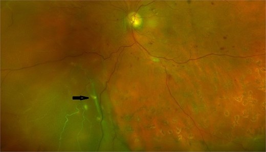
Color fundus photograph of the left eye showing retinal detachment with a cylindrical dexamethasone implant in the subretinal space, indicated by an arrow. The remaining retina shows proliferative diabetic retinopathy changes with pan retinal photocoagulation scars.
After informed consent, the patient was scheduled for pars plana vitrectomy with retinal detachment repair. Given prior reports describing potential spontaneous biodegradation of the implant and the absence of established management guidelines, an initial strategy of leaving the implant in situ was considered.
A 25-gauge pars plana vitrectomy was performed with chandelier-assisted bimanual illumination. During core vitrectomy, the subretinal dexamethasone implant was observed to migrate into the vitreous cavity through the pre-existing retinal break. In light of the risk of postoperative anterior migration with subsequent corneal endothelial toxicity, the surgical plan was revised intraoperatively to pursue implant removal.
Perfluorocarbon liquid (perfluoro-n-octane) was instilled to stabilize and protect the posterior pole, thereby minimizing the risk of mechanical trauma to the retina and optic nerve during implant manipulation. The implant was engaged using 25-gauge end-gripping forceps (Alcon Grieshaber AG, Schaffhausen, Switzerland). Attempts at en bloc removal using a low cut-rate vitreous cutter were unsuccessful due to fragmentation of the implant.
A bimanual technique was subsequently employed. Individual fragments were sequentially regrasped with forceps, advanced toward the cutter port, and aspirated under controlled low-flow settings. Perfluorocarbon liquid provided countertraction and maintained posterior pole stability throughout the maneuver. Following removal of the larger fragments, meticulous peripheral vitreous shaving was performed under bimanual visualization, and residual particulate material at the vitreous base was aspirated to minimize the risk of retained fragments.
After completion of the vitrectomy, a fluid–air exchange was performed, and subretinal fluid was drained through the existing retinal break. Endo laser photocoagulation was applied to the break, followed by gas tamponade with 14% perfluoro propane (C3F8). Sclerotomies were closed with 6-0 plain gut sutures, and subconjunctival antibiotics were administered (see Additional file). Postoperative course was uneventful and BCVA was 20/20 after resolution of gas bubble at 90 days.
Discussion
Reports of subretinal localization of a dexamethasone 0.7-mg intravitreal implant (Ozurdex) are exceedingly limited, and previously described cases have largely involved eyes with an intact neurosensory retina managed conservatively [6]. In contrast, the concurrence of retinal detachment with intraoperative migration of the implant from the subretinal space into the vitreous cavity, as observed in the present case, represents an uncommon and insufficiently characterized clinical scenario. The intraoperative finding of a free-floating implant over the posterior pole following migration through a pre-existing retinal break underscores the dynamic behavior of the implant in the absence of vitreous support.
The pathogenesis of subretinal implant localization remains incompletely defined. Prior reports have implicated suboptimal injection technique, including excessive obliquity of the applicator or injection at a distance greater than 4 mm posterior to the limbus, potentially resulting in direct retinal impact or penetration [6]. Alternatively, in the current case, it is plausible that a pre-existing retinal detachment permitted secondary migration of the implant into the subretinal space. Conversely, inadvertent retinal injury at the time of injection may have created a retinal break, subsequently leading to rhegmatogenous detachment with subretinal entrapment of the implant. These mechanisms are not mutually exclusive and likely reflect the interplay between injection dynamics and altered vitreoretinal anatomy.
Management of subretinal dexamethasone implants remains undefined. In previously reported cases without associated retinal detachment, conservative observation has been described, with gradual biodegradation of the implant and minimal long-term sequelae [6]. However, the presence of concurrent retinal detachment introduces additional considerations that favor surgical intervention. In the present case, intraoperative migration of the implant into the vitreous cavity prompted removal, given the recognized risk of postoperative anterior chamber migration and subsequent corneal endothelial toxicity. At present, there are no established guidelines addressing implant management in the context of retinal detachment or intraoperative migration, and surgical decision-making must therefore be individualized.
Several techniques have been described for removal of intravitreal corticosteroid implants. These include direct grasping with micro-forceps and extraction through a sclerotomy, as well as bimanual approaches combining forceps manipulation with vitreous cutter-assisted aspiration [7, 8]. The biodegradable polymer matrix of the dexamethasone implant predisposes it to fragmentation during manipulation, particularly when attempts are made at en bloc removal. In our case, fragmentation occurred upon attempted grasping, necessitating a controlled bimanual technique. Sequential regrasping of fragments with end-gripping forceps, combined with low-flow aspiration using the vitreous cutter under perfluorocarbon stabilization, allowed safe and complete removal. Although technically demanding and time-intensive, this approach provides effective control of mobile fragments and minimizes the risk of retinal injury.
In conclusion, subretinal lodgment of a dexamethasone intravitreal implant is a rare iatrogenic complication. Careful attention to injection technique, including appropriate entry site and angle, as well as thorough peripheral retinal evaluation before and after injection, is essential to mitigate risk. In the setting of retinal detachment with intraoperative migration of the implant into the vitreous cavity, surgical removal should be strongly considered. This case highlights the utility of perfluorocarbon-assisted bimanual techniques for safe retrieval in complex intraoperative scenarios.
Conflicts of interest
No authors have any proprietary interest in this study.
Funding
None declared.
Data availability
The dataset supporting the conclusions of this article is included within the article (and its additional file).


