


 https://orcid.org/0000-0002-5286-3532

Abstract
Neuroendocrine carcinomas of the colon are aggressive and rare tumors that often present locally advanced and/or with distant metastases at diagnosis. Their incidence is reported to be <1%. Surgery plays a major role as the initial treatment for locally advanced disease, followed by adjuvant chemotherapy, since most of the cases present in the advanced stage. Despite this, the overall prognosis is reported to be worse, with a median survival of 9 to 14 months. This is a case of an aggressive large cell neuroendocrine carcinoma of the right colon treated with upfront surgery followed by adjuvant chemotherapy.
Introduction
Neuroendocrine tumors (NET) account for < 1% of all colorectal malignancies [1]. A rising incidence of NET of the colon, and especially of the rectum, has been reported in the last two decades [2]. Their high level of differentiation, low to moderate mitotic rate, and proliferative index (Ki–67) of <20% result in better treatment outcomes and favorable patient prognoses. On the contrary, neuroendocrine colorectal neoplasms with high-grade morphology, >20 mitoses / 2 mm2, and a high proliferation index of over 20% are referred to as neuroendocrine carcinomas (NEC) and have a far worse prognosis [3].
The reported incidence of high-grade colon neuroendocrine carcinoma is 0.6% of all colorectal carcinomas [4]. The poor prognosis for this cancer is reflected in the reported median overall survival of 9 months. The mainstay for non-metastatic colon NEC is surgery followed by adjuvant chemotherapy [1]. We present a case of an aggressive large cell type NEC of the caecum in a male patient treated with right hemicolectomy and adjuvant chemotherapy with intra-abdominal and early retroperitoneal recurrence 8 months after surgery, despite proper oncologic R0 resection. Written informed consent was obtained from the patient.
Case report
A 70-year-old male patient with previously diagnosed adenocarcinoma of the right colon was admitted to our hospital for treatment. No comorbidities were reported. The only complaint was occasional pain in the lower right abdominal quadrant. A palpable mass in the right iliac fossa was present on physical examination. Abnormal laboratory findings on admission day encountered the level of haemoglobin of 113 g/L (120–165 g/L), thrombocyte count of 456 × 109/L (150–390 109/L), C-reactive protein value of 66.5 mg/L (0–5 mg/L), serum total protein level of 51.7 g/L (64–83 g/L), serum albumin level of 28.8 g/L (35–50 g/L), serum iron level of 4 μmol/L (11.6–31.3 μmol/L), and serum D-dimer value of 1609 ng/ml (0–500 ng/ml). Abdominal computed tomography (CT) showed a mass in the right colon and mesenteric lymph nodes along the ileocolic vessels, with a maximum diameter of 25 mm (Fig. 1). Exploratory laparotomy revealed a tumor of the cecum, and open right hemicolectomy with primary ileum-transverse anastomosis was performed. The postoperative period was uneventful, and the hospital stay lasted 5 days.
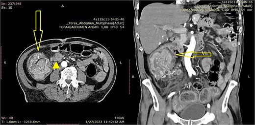
Abdominal CT (axial scan) from January 2023 showing a large tumor mass (arrow) with a significantly enlarged lymph node (arrowhead) (a - axial scan, b - coronal scan).
The macroscopic pathology report described the presence of a tumor in the caecum at the level of the ileocecal valve with a measured length of 10 cm and a diameter of 7 cm. It engaged 3/10 of the colon circumference with infiltration depth up to the subserosa. Proximal and distal resection margins from the tumor were measured at 19 and 17 cm, respectively. Eighteen lymph nodes were isolated, and a metastatic deposit was detected in 6 of them. Extramural lymphovascular invasion was confirmed.
Histopathological analysis revealed the presence of poorly differentiated neuroendocrine carcinoma with high histologic and nuclear grade, built from round cells with round nuclei arranged in organoid structures. There were 34 mitoses present on 10 High Power Fields (Fig. 2).
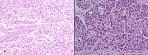
Haematoxylin / eosin stain (a – 100× magnification, b – 400× magnification).
Additional immunohistochemistry stain showed strong positivity for cytokeratin (CKE) AE1/AE3 and a slight positive signal for Synaptophysin. The tumor population was negative for Chromogranin, NSE, CD56, CK20, and CDX2. The proliferative index was >20% (Fig. 3). The final pathologic stage according to the Union for International Cancer Control (UICC – 8th edition) was pTNM = pT3, pN2а, pM0, pR0, pL1, pV1, G3, NG3, Stage IIIВ. Molecular analysis revealed a microsatellite-stable BRAF Wild Type of cancer.
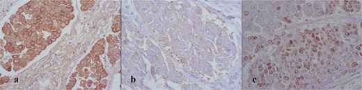
Immunohistochemistry stain showing positivity for CKE AE1/AE3 (400×) (a), synaptophysin (400×) (b), and Ki-67 proliferative index >20% (400×) (c).
The patient was referred to an oncologist, where an adjuvant Cisplatin-Etoposide protocol was given. Two months after surgery, a CT scan showed the presence of paraaortal, paracaval, and mesenteric enlarged lymph nodes with a maximal diameter up to 21 mm without evident signs of local recurrence.
The effect of the given adjuvant chemotherapy regimen was evaluated with a 18F-FDG PET/CT scan (8 months post-op). It revealed the presence of multiple metabolically active enlarged mesenteric and retroperitoneal lymph nodules (SUV max = 15.3, d = 28 mm), which were described to have malignant PET/CT characteristics (Fig. 4).
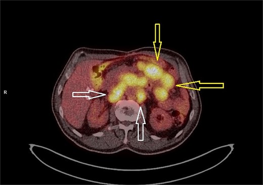
18F-FDG PET/CT scan from September 2023 showing metabolic activity in the retroperitoneal (white arrows) and mesenteric (yellow arrows) lymph nodes.
The patient was scheduled for a physical exam. A palpable abdominal mass was present. An abdominal CT scan showed the presence of multiple lymph node packages spreading caudally from the celiac trunk up to the pelvis. Multiple intra-abdominal lesions with a maximal diameter of up to 45 mm were also detected, producing mass effect on the intestines. A small amount of intra-abdominal fluid was detected in the retrovesical space (Fig. 5).
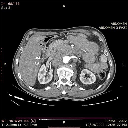
Abdominal CT (axial scan) from September 2023 showing massive retroperitoneal lymphadenopathy and intra-abdominal lesions.
The oncologist initiated 6 cycles of second-line chemotherapy with leucovorin, 5-fluorouracil, and irinotecan (FOLFIRI protocol) for 4 months. Due to the disease progression despite the second–line therapy, the oncologist initiated a third-line therapy with capecitabine and temozolomide (CAPTEM protocol). However, the patient's condition continuously deteriorated, and the patient passed away 19 months after surgery.
Discussion
This case presents the aggressive behavior of poorly differentiated NEC of the cecum. NEC’s are found in the colon in less than 1% of all colorectal tumors, with the higher rates reported in Europe and North America [5]. In the series of Muğlu et al. NEC was found in the colon in 4% of the patients with extrapulmonary NEC [6]. According to Bernick et al., the incidence of LCNEC of the colon is reported to be 0.2% [7]. Most of the extrapulmonary NECs are non-hormone-secreting tumors. Due to the lack of early symptoms, many patients present with advanced- or late-stage disease [6].
According to histology, a clearly defined neuroendocrine pattern includes uniform, regular cells with round or oval nuclei, salt-and-pepper chromatin, and moderate eosinophilic granular cytoplasm. In organoid structures, cancer cells are organized in clusters, trabecular, or insular formations. Nuclear pleomorphism may be observed in certain instances (endocrine atypia), but it does not correlate with tumor aggressiveness. There can be little mucin release. The grading system for NET relies on the rate of proliferation (mitoses and Ki-67 index) as follows: Grade 1 or low grade (G1): mitoses <2 per 2 mm2 and Ki-67 index <3%; Intermediate grade or grade 2 (G2): mitotic figures 2–20 per 2 mm2 or Ki-67 index 3–20%; High grade or grade 3 (G3): mitotic count >20 per 2 mm2 or Ki-67 index >20%. Mitotic count must be assessed in a 2 mm2 hotspot region (approximately equal to 10 high-power fields using a 40× objective lens). The Ki-67 index must be evaluated in at least 500 cells within the hotspot areas. In cases of discrepancy between the mitotic index and the Ki-67 index, the higher value should be used for classification. NEC morphology includes a large-cell type (LCNEC) and a small-cell type (SCNEC). LNEC shows organoid cell structures larger than those found in small cell carcinoma, exhibiting nuclear pleomorphism and hyperchromasia, prominent nucleoli, numerous mitotic figures, and tumor necrosis. SNEC is similar to pulmonary small cell carcinoma, characterized by clusters and aggregates of small oval cells with scant cytoplasm, hyperchromatic nuclei with stippled chromatin, nuclear molding and peripheral palisading, active mitosis, apoptotic cells, necrosis, and invasion of blood vessels [3].
Surgery in LCNEC offers certain survival in patients despite the overall poor prognosis. In selected cases with Microsatellite Instability-High (MSI-high) status, the addition of immunotherapy and molecularly targeted therapies may be beneficial. The standard first-line adjuvant chemotherapy regimen is platinum-based. Second-line and third-line chemotherapy are reported to result in partial and no response, respectively [6]. The benefits of adjuvant chemotherapy were questioned by Wu et al. In their study on 318 patients, they showed no benefits in cancer-specific survival and overall survival if etoposide or 5-FU regimens were added to the surgery in Stage I-III NEC [8]. In this case, the patient presented with a locally advanced cancer treated with upfront surgery followed by first-line, and then second and third-line therapy, without achieving a stable disease.
Data on the neoadjuvant treatment in patients with colon NEC is scarce. However, according to the European Neuroendocrine Tumor Society 2023 guidance paper for colorectal neuroendocrine tumors, this treatment approach might be reasonable in highly selected patients with locally advanced disease [2]. In addition to this, even a complete pathological response is reported in a case with a localized small cell colon carcinoma [9].
The reported median survival rates are between 9.0 and 14.7 months [1, 7, 10, 11]. Yet, certain series report a prolonged survival [12, 13]. In this report, the patient’s survival is slightly above the previously reported ranges. Factors influencing the survival in patients with NEC are initial presentation without metastases, presence of adenocarcinoma component in the tumor, and response to the given chemotherapy [11].
Conclusion
Colon NEC remains a highly aggressive malignancy despite the combined treatment modalities as presented in this case report. A priority might be given to the neoadjuvant chemotherapy in selected cases.
Conflicts of interest
None declared.
Funding
None declared.
References
Muğlu H, Sünger E, Mıldanoğlu MM, et al. Clinicopathological characteristics of extrapulmonary neuroendocrine carcinomas: treatment responses and survival outcomes: single-center experience.


