


Abstract
Pectus excavatum (PE) is the most common congenital chest wall deformity but its association with atrial fibrillation (AF) remains underrecognized. We present a case where surgical correction of PE resulted in complete resolution of AF, highlighting the role of structural compression in lone AF and other arrhythmias. A 25-year-old male with PE presented with recurrent symptomatic AF without prior cardiovascular disease. Despite initial treatment with metoprolol and apixaban, episodes persisted. Chest CT demonstrated severe deformity (Haller index 5.6) and right atrial compression. Echocardiography showed a reduced ejection fraction (40%–45%). He underwent a Nuss procedure with dual bars and intercostal nerve cryoablation, resulting in immediate intraoperative relief of compression. There was no recurrence of AF at 9 months follow up. Severe PE may contribute to lone AF through mechanical compression. Surgical correction can provide symptom resolution in appropriate patients.
Introduction
Pectus excavatum (PE) is the most common congenital chest wall deformity, affecting 1 in 800–1000 live births [1]. PE can cause compression of the right ventricle, right atrium, and outflow tract, leading to dyspnea, exercise intolerance, tachycardia, and reduced stroke volume [2]. Atrial fibrillation (AF), typically associated with aging and cardiovascular disease, results in significant morbidity. Lone atrial fibrillation, defined as AF in patients younger than 60 without structural heart disease or traditional risk factors, has been associated with significant cardiac abnormalities, including mitral valve prolapse and other arrhythmias [2].
The Haller Index is commonly used to define the morphological severity of PE, which is calculated by dividing the maximum transverse diameter of the chest on computed tomography (CT) by the minimum distance from the sternum to the vertebral column [2]. Evidence of functional impairment on pulmonary function tests and cardiopulmonary exercise testing drives the decision to correct PE, which is usually done through the minimally invasive Nuss procedure [3]. Select case studies have demonstrated reversal of electrocardiogram (EKG) abnormalities, showing improvements in exercise tolerance and elimination of symptoms post-correction [4, 5]. We present the case of lone AF in a young adult with severe PE where surgical correction resulted in resolution of AF, highlighting a potential reversible etiology.
Case report
A 25-year-old male presented to the emergency department after feeling lightheaded when standing and was found to be in hemodynamically stable AF. Previous medical history included untreated PE, which was characterized as cosmetic, and intermittent exertional dyspnea and chest discomfort. There was no family history of connective tissue disorders or arrhythmias. After initiation of medical therapy, he had repeated episodes of asymptomatic AF and was evaluated for ablation. Echocardiography demonstrated mid-range reduced ejection fraction (40%–45%) with no intrinsic structural abnormalities. Pulmonary function testing showed mild obstructive physiology consistent with asthma, and he was initiated on budesonide. On physical exam, there was diffuse sternal depression (“Grand Canyon type”) and mild costal flaring. PE (Haller index 5.6) and severe right heart compression were seen on chest CT, raising suspicion that sternal compression contributed to his AF and reduced ejection fraction (Fig. 1). Mechanical cardiac compression from PE was considered a contributing etiology, and a surgical referral was made.
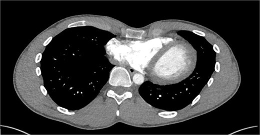
CT of chest prior to surgery demonstrating significant right atrio-ventricular compression due to PE with Haller index of 5.6.
Four months after initial presentation, the patient underwent thoracoscopic PE repair with the Nuss procedure using dual titanium bar placement and bilateral intercostal nerve cryoablation. Severe sternal depression with rightward torsion was noted, extending from the chondromanubrial junction to the xyphoid. Two 32 cm titanium bars were placed using thoracoscopic guidance due to extensive sternal depression and a more rigid adult chest wall. Right atrial compression relief was noted after sternal elevation (Figs 1 and 2).
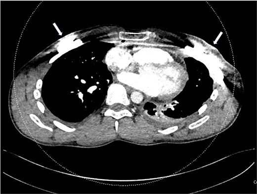
Chest imaging after surgery demonstrating resolved compression of the right atrium and ventricle. The titanium pectus bars used to correct the PE are seen as artifact laterally (arrows).
The patient was discharged on POD1. On POD2, he presented to the emergency department (ED) with cough, low-grade fevers, and dyspnea attributed to inadequate pain control and atelectasis. Chest X-ray (CXR) revealed trace bilateral apical pneumothoraces, trace bilateral pleural effusions, and basilar atelectasis. EKG demonstrated normal sinus rhythm. His symptoms improved with optimized analgesia. A CT angiogram of the chest was obtained, which demonstrated post-surgical free air in the mediastinum but no pulmonary embolism (Fig. 3). At 1-month follow-up, the patient reported resolution of dyspnea and wheezing. He has had no recurrence of cardiac issues or AF postoperatively at 9 months of follow-up. The authors confirm that patient consent forms have been obtained for this article.
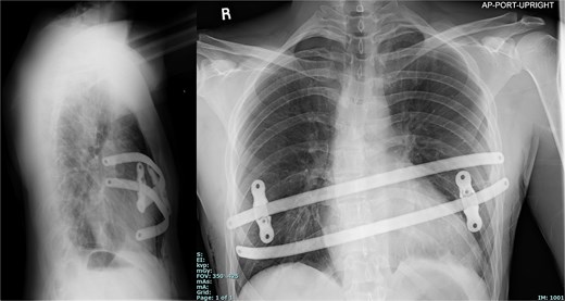
CXR demonstrating bar placement post-repair.
Discussion
This case report supports an underrecognized association between severe PE and AF due to mechanical cardiac compression. While AF is typically attributed to age-related remodeling or cardiovascular disease, structural compression of the heart represents a potentially reversible cause in young patients with PE. In young patients with severe PE, evidence of chamber compression and AF in the absence of another clear etiology, surgical correction of pectus should be considered as attempts at more traditional therapies such as antiarrhythmic drugs or ablation fail to address what is ultimately of anatomical origin. While previous cases have demonstrated the presence of various arrhythmias and their association with PE, the outcome of surgical intervention has not been described in detail. This case illustrates how surgical intervention can improve cardiac function in severe PE cases and provide cosmetic and pulmonary relief.
This case also demonstrates the complexity of PE correction in an adult due to increased chest wall rigidity, requiring a longer operative time and placement of two titanium bars instead of one. It highlights the importance of early intervention for PE to prevent morbidity and lower operative complications. Although the post-operative findings (mild atelectasis and pneumothoraces) in this patient were expected after surgery, pain control was a challenge.
This case demonstrates AF in an otherwise structurally normal heart, supporting the decision to pursue PE correction first. Compression and subsequent reduced stroke volume can evolve to compensatory resting sinus tachycardia, which can trigger lone AF. Clinicians should maintain a high index of suspicion for PE-related cardiac effects in young patients presenting with unexplained AF, particularly when imaging demonstrates right-sided chamber compression. Early recognition may prevent unnecessary interventions and allow for more definitive treatment. A limitations of the study is that it is a single case report, which limits the generalizability of the findings. Further prospective studies are needed to explore the pathophysiology of AF development in the setting of PE and understand the efficacy of PE correction in the treatment of AF.
Author contributions
R.L.: literature review, data interpretation, drafting of article, revision of manuscript; S.L.: literature review, data interpretation, drafting of article, revision of manuscript; P.H.: data interpretation, revision of manuscript; and F.D.: concept design, data interpretation, revision of manuscript, approval of article
Conflicts of interest
None declared.
Funding
No funding was received for this work.
Data availability
The data that support the findings of this study are available from the corresponding author, F.D., upon reasonable request.
Informed consent statement
The patient’s informed written consent was obtained for publication of study data.


