


 https://orcid.org/0009-0003-5333-5875

Abstract
Capsule endoscopy is an established diagnostic modality for small bowel evaluation, particularly in Crohn’s disease. Capsule retention remains its most significant complication, especially in patients with stricturing disease. We report two cases of asymptomatic capsule retention in patients with established Crohn’s disease. In both cases, cross-sectional imaging failed to predict clinically significant stenosis. Capsule retention was confirmed radiologically after 2 weeks. Corticosteroid therapy did not result in capsule passage, and both patients ultimately required surgical intervention. Surgical management allowed capsule retrieval and definitive treatment of underlying strictures through bowel resection and stricturoplasty. These cases highlight the importance of appropriate pre-procedural risk stratification and multidisciplinary management. Routine use of cross-sectional imaging and, when available, patency capsule testing is strongly recommended in patients at risk of small bowel stenosis.
Introduction
Capsule endoscopy has revolutionized the investigation of small bowel pathology and is widely used for obscure gastrointestinal bleeding and Crohn’s disease evaluation [1, 2]. In Crohn’s disease, it allows early detection of mucosal lesions, assessment of disease extent, and evaluation of mucosal healing [3].
However, capsule retention remains the most significant complication of the procedure. It is defined as the presence of the capsule in the gastrointestinal tract for more than 2 weeks or requiring medical, endoscopic or surgical intervention for removal [4]. The overall retention rate is ~1%–2%, but it increases substantially in patients with established Crohn’s disease, reaching up to 13% in some series [5, 6].
We report two cases of asymptomatic capsule retention in patients with Crohn’s disease requiring surgical management.
Case report
Case 1
A 47-year-old man with terminal ileal Crohn’s disease diagnosed in 2006 and treated intermittently with azathioprine presented with Koenig’s syndrome and constipation. Ileo-colonoscopy was normal. Computed tomography (CT) enterography showed multifocal ileal wall thickening without definite stenosis. Capsule endoscopy (PillCam SB3) revealed aphthous jejunal ulcerations and an ulcerated distal jejunal stricture (Fig. 1).
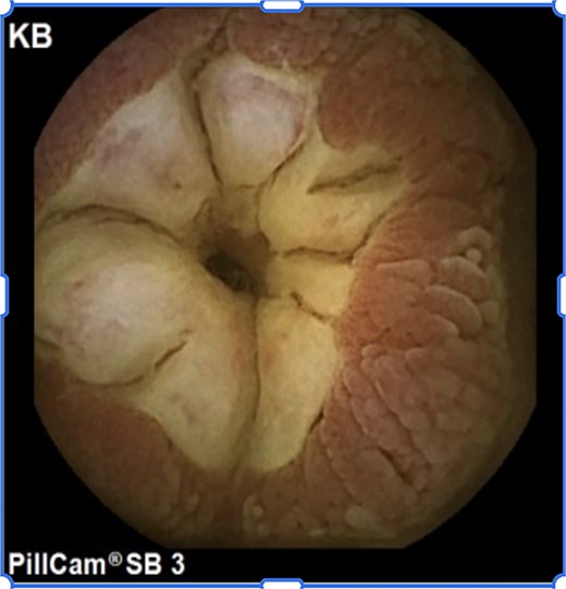
Capsule endoscopy image showing ulcerated jejunal stricture (Case 1).
The capsule was not expelled after 2 weeks. Abdominal radiography and CT confirmed jejunal retention (Fig. 2). The patient remained asymptomatic. Corticosteroid therapy was initiated but failed to induce capsule passage.
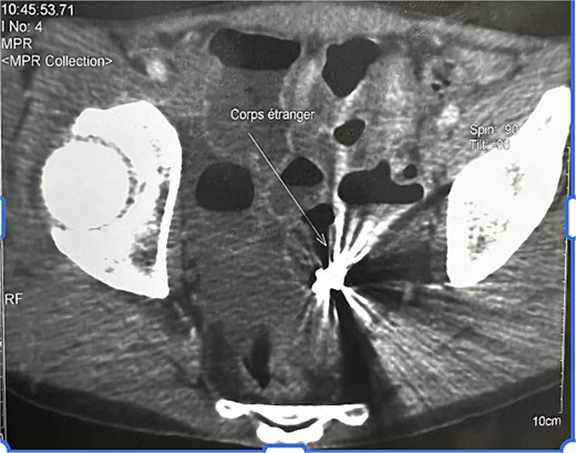
Abdominal CT demonstrating jejunal capsule retention (Case 1).
Surgical exploration performed 3 months later revealed multiple small bowel strictures with upstream dilatation. A 40 cm segment containing the capsule was resected, and a temporary ileostomy was performed.
Case 2
A 35-year-old man with Crohn’s ileitis diagnosed in 2022 and treated with infliximab and azathioprine was investigated for iron-deficiency anemia. Upper endoscopy and ileo-colonoscopy were normal. Magnetic resonance enterography demonstrated inflammatory thickening of the distal ileum without clear stenosis. Capsule endoscopy (CapsoCam) was performed (Fig. 3).
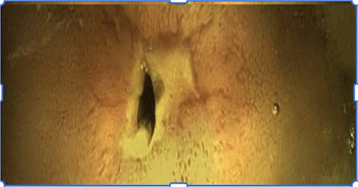
Capsule endoscopy image showing ileal fibrotic stricture (Case 2).
The capsule was not expelled after 2 weeks. Abdominal radiography and CT confirmed proximal ileal retention (Fig. 4). The patient remained asymptomatic. Corticosteroids were administered without success.
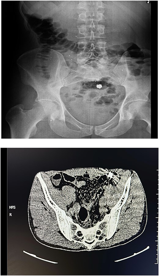
Abdominal X-ray and CT confirming capsule retention in proximal ileum (Case 2).
Surgical exploration revealed multiple non-occlusive strictures. Management included resection of one stenotic segment, stricturoplasty of another, and dilation of two additional strictures. The capsule was retrieved successfully. Analysis confirmed an ulcerated ileal stricture at the site of retention.
Discussion
Capsule endoscopy plays a central role in the evaluation of small bowel Crohn’s disease due to its high sensitivity for superficial mucosal lesions [2, 3]. Nevertheless, capsule retention represents its principal limitation and may necessitate surgical intervention [4].
Retention is strongly associated with stricturing disease. A meta-analysis reported retention rates of ~2% overall and significantly higher rates in established Crohn’s disease [5]. Importantly, retention may occur in asymptomatic patients, as demonstrated in both of our cases [6].
Pre-procedural cross-sectional imaging such as CT or magnetic resonance enterography is recommended to assess stenosis [7]. However, imaging may underestimate the functional significance of inflammatory or short strictures. In our patients, imaging demonstrated inflammatory thickening but failed to predict retention.
The patency capsule is an effective tool to reduce retention risk, with high sensitivity and negative predictive value for detecting significant stenosis [8]. Current guidelines recommend its use in patients with known Crohn’s disease or suspected strictures [9]. Unfortunately, patency capsule testing was unavailable in our institution.
Management of capsule retention depends on symptoms and underlying pathology. In asymptomatic patients, medical therapy may promote passage if inflammation predominates [10]. Device-assisted enteroscopy may allow endoscopic retrieval [11]. However, when retention persists or strictures are fibrotic, surgery remains definitive.
In both cases, surgery enabled capsule retrieval and treatment of stricturing disease through resection and stricturoplasty. These cases emphasize the importance of multidisciplinary decision-making and appropriate risk stratification before capsule endoscopy.
Conflicts of interest
The authors declare no conflict of interest.
Funding
None declared.


