


Abstract
Spinal anesthesia (SA) is preferred for cesarean delivery but poses technical challenges in patients with spinal deformities or cerebrovascular disorders. This report describes the successful use of ultrasound-assisted SA in a parturient with surgically corrected scoliosis and moyamoya disease. A 28-year-old female at 37 weeks of gestation with history of surgery for scoliosis (post-thoracolumbar instrumentation) and moyamoya disease underwent elective cesarean section. Severe spinal curvature, metallic implants, and scarring obscured anatomical landmarks. Preprocedural ultrasound identified the L3–L4 interspace, enabling single-attempt SA with optimal sensory blockade (T8–T10). No complications occurred. Ultrasound guidance enhances SA success in complex cases by overcoming anatomical challenges, ensuring hemodynamic stability, and minimizing procedural risks. This technique warrants broader adoption in patients with spinal deformities and comorbid conditions.
Introduction
Spinal anesthesia (SA) remains the gold standard for cesarean delivery due to its maternal and fetal safety profile, rapid onset, and reduced systemic toxicity risk compared to general anesthesia [1, 2]. However, anatomical abnormalities—such as scoliosis—increase the likelihood of failed punctures and complications [3]. Concurrent cerebrovascular conditions like moyamoya disease further necessitate precise hemodynamic control, favoring SA to avoid cerebral hypoperfusion or hyperventilation-induced ischemia [4, 5]. Ultrasonography addresses these challenges by enabling real-time visualization of spinal structures, improving first-attempt success rates in complex cases [3, 6].
Case presentation
A 28-year-old female at 37 weeks and 3 days of gestation presented for elective cesarean section. Her medical history included scoliosis corrected via thoracolumbar instrumentation in 2011 and moyamoya disease treated with left superficial temporal artery–middle cerebral artery bypass surgery in December 2022. Residual scoliosis, spinal hardware, and surgical scarring obscured surface landmarks. Physical examination revealed persistent thoracolumbar curvature (Cobb angle not quantified), poor alignment of spinous processes, and scarring over the surgical site (Fig. 1). Neurological evaluation showed no deficits. Preoperative lumbar spine radiographs confirmed residual scoliosis and instrument placement (Fig. 2). Magnetic resonance angiography demonstrated stable moyamoya vasculature without active stenosis.
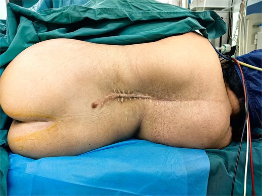
Patient positioning demonstrating spinal curvature.
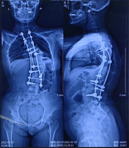
Radiographs showing thoracolumbar instrumentation and residual scoliosis.
After obtaining informed consent, ultrasound-assisted SA was planned. Using a Mindray UMT-400 ultrasound with a C5-2S probe, the L3–L4 interspace was identified by tracing transverse processes from the sacrum. Skin-to-subarachnoid depth (6.5 cm) and needle trajectory were measured. Guided by the premeasured landmarks, the dural puncture was successfully achieved with a 25-gauge spinal needle on the first attempt. 1.4 mL of 0.75% bupivacaine was injected and the sensory blockade was titrated to T8–T10. Hemodynamics remained stable (MAP 70–80 mmHg), and the cesarean section proceeded uneventfully. The patient reported no post-dural puncture headache or neurological symptoms at 24-h follow-up. The patient expressed satisfaction with the procedure’s efficiency and postoperative pain control.
Written informed consent was obtained from the patient for both the procedure and publication of anonymized data. Ethical approval was waived per institutional policy for retrospective case reports.
Discussion
This case highlights the utility of ultrasound-assisted SA in a parturient with dual complexities of scoliosis and moyamoya disease. Scoliosis disrupts spinal anatomy, increasing the risk of multiple punctures and complications such as nerve injury [7, 8]. Ultrasound mitigates these risks by visualizing interspaces and optimizing needle angles, as demonstrated by a first-attempt success rate of 87.5% in similar populations [9, 10]. Moreover, a randomized controlled trial revealed that ultrasound-assisted neuraxial anesthesia markedly decreases the incidence of complications such as lower back pain and headache [11].
Approximately 70% of pregnant women with moyamoya disease would undergo cesarean sections to mitigate risks of cerebral hemorrhage or infarction [12]. For moyamoya disease, SA’s hemodynamic stability is critical to prevent cerebral ischemia [5]. General anesthesia risks hyperventilation-induced vasospasm, whereas SA minimizes blood pressure fluctuations, aligning with retrospective evidence favoring SA in moyamoya parturients [5, 12].
This case aligns with prior studies advocating ultrasound for challenging neuraxial anesthesia [11, 13]. A randomized trial by Chin et al. [14] reported a 95% success rate with ultrasound in patients with difficult anatomy, compared to 68% with landmark techniques. Our experience reinforces these findings, emphasizing ultrasound’s role in reducing procedural time and complications.
Conclusion
Ultrasound-assisted SA is a safe, efficient, and patient-centered approach for cesarean delivery in parturients with spinal deformities and comorbid cerebrovascular disease. Its integration into clinical practice should be prioritized to enhance procedural success and maternal outcomes.
Conflict of interest statement
The authors report no conflicts of interest.
Funding
The work was funded by the Institutional project (2023LC2352, XJZT24LY15).
References
Author notes
Yongzhou Jiang, Yonghui Wang and Fahan Wang contributed the same to the manuscript.
