


Abstract
This case report presents a preterm infant (36 weeks) with gastroschisis, successfully treated using a two-stage surgical approach. The newborn exhibited evisceration of the stomach and small intestine through a small umbilical defect, with significant edema but no additional anomalies. Due to the severity of the edema and limited abdominal capacity, a staged repair was planned. The first stage involved repositioning the stomach and duodenum into the abdominal cavity. In the second stage, the remaining bowel was placed in a sterile sac suspended above the incubator, allowing gradual reduction over 2 days. Enteral nutrition was initiated after 1 week. The approach resulted in a smooth recovery without complications, suture failure, or incisional hernia. This case highlights the importance of meticulous surgical planning in improving outcomes for neonates with complex conditions.
Introduction
Gastroschisis (GS) is a paraumbilical defect of the anterior abdominal wall, resulting in prenatal intestinal herniation into the amniotic space, leading to varying degrees of intestinal exposure and requiring early postnatal care [1]. The etiology remains largely unknown, though young maternal age is a significant risk factor [2]. Over the past three decades, GS incidence has increased, with regional variation, and it occurs in approximately 1 in 1953 births globally [2]. Recent studies report GS prevalence between 1.1 and 5.1 per 10 000 live births [1]. Complicated GS, involving intestinal atresia, perforations, necrosis, and volvulus, is associated with a poorer prognosis than simple GS, which typically lacks intestinal complications. Prenatal diagnosis is made using ultrasonography, including 3D ultrasound in some cases [2]. Surgical interventions aim to close the abdominal defect, prevent abdominal compartment syndrome, and safely reduce the herniated viscera, either through immediate or staged approaches, such as the use of a prosthetic silo for gradual reduction [3, 4].
Case presentation
A 36-week preterm neonate with GS was referred to our hospital. Upon admission, significant evisceration of the stomach and small bowel was observed, accompanied by notable edema (Fig. 1). The herniated contents could not be reintroduced immediately due to their size. Physical examination revealed no signs of macrosomia or other anomalies. A two-stage surgical approach was planned: first, the stomach and duodenum were returned to the abdomen, followed by covering the herniated bowel with a sterile sac (Fig. 2), suspended from the incubator ceiling to function as a makeshift silo. Over the next 2 days, gradual reduction of the herniated bowel occurred within this sac (Fig. 3). One week post-surgery, enteral feeding was initiated without complications, and the patient was discharged in stable condition, with no signs of incisional hernia or suture failure.
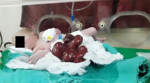
Evisceration of the stomach and small bowel.
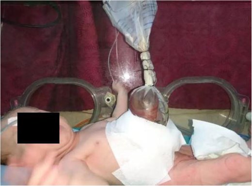
The gastroschisis contents were covered with a sterile sac.
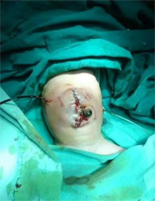
A careful and staged reduction of the herniated bowel was successfully conducted.
Discussion
Gastroschisis involves evisceration of abdominal organs, often including the intestines, without a protective membrane. The defect is usually located to the right of the umbilicus and is typically small, about 4 cm in diameter [5]. In our case, multiple organs, including the intestines, duodenum, stomach, liver, and spleen, were involved. The incidence of GS has increased in recent years, with a prevalence ranging from 0.4 to 3 per 10 000 births. Most diagnoses occur between 14 and 30 weeks of gestation, and deliveries typically occur between 34.8 and 38.2 weeks. The male-to-female ratio ranges from 1.0 to 1.4 [6].
The development of GS involves the abnormal exteriorization of the intestine, beginning in the first trimester and continuing into the third trimester [1]. Prenatal ultrasound is highly effective for diagnosing GS, with a fetal identification rate exceeding 90% when combined with maternal serum alpha-fetoprotein screening. The morphology of the defect helps differentiate GS from omphalocele [1]. Approximately 95% of GS cases are isolated defects, though some associated anomalies may occur, particularly non-duodenal intestinal atresia (10%) [7]. Mortality is higher in cases with associated anomalies, especially those involving intestinal complications [6]. Complications, such as delayed enteral feeding, extended parenteral nutrition and prolonged hospital stays, are common [8].
In our case, no significant complications arose aside from partial liver herniation. Liver herniation is a known risk factor for poor outcomes, with a high mortality rate in cases of severe herniation [9]. Prenatal detection allows for delivery in a specialized setting, where the patient can be managed by a multidisciplinary team [10]. Timing of delivery is important; some studies suggest avoiding deliveries before 35 weeks to reduce risks [11]. Overall, survival rates for isolated GS cases exceed 90%, though some neonates experience substantial long-term complications [12].
Postnatal care in GS involves managing fluid loss, heat, and potential respiratory risks, with nasogastric tube insertion for airway protection and fluid compensation. Antibiotic therapy is essential for preventing wound infections and catheter-related infections [13]. In our institution, ampicillin and gentamicin are routinely prescribed after silo placement, especially for delayed closure procedures.
Defect closure can be immediate or delayed, with a prosthetic silo used for gradual reduction in certain cases. Primary closure is preferred to prevent mechanical injury, wound infections, and vascular compromise, though it is not always feasible [12]. Silo placement, which avoids suturing, is an alternative that can be performed on an awake neonate [3]. In our case, the patient underwent a two-stage procedure, with the first stage involving repositioning the stomach and duodenum and the second stage involving the gradual reduction and final closure.
Conclusion
This case report highlights the successful management of GS in a preterm neonate through a two-stage surgical approach. The use of a sterile sac as a protective silo for gradual reduction allowed for optimal recovery and safe closure of the abdominal defect. The patient’s postoperative course was favorable, with no complications or signs of incisional hernia. This case supports the efficacy of the two-stage surgical approach for managing complex GS cases and underscores the need for continued research to refine surgical techniques and improve long-term outcomes in affected neonates.
Acknowledgements
We hope to thank SMSR Team Lab for their efforts and for bringing our team together.
Author contributions
M.S., M.A., H.A., B.S., H.A., A.M., A.B., M.F., F.A., A.O. wrote part of the manuscript. All authors approved the final manuscript.
Conflict of interest statement
No conflict of interest.
Funding
None declared.
Data availability
The data that support the findings of this study are available from the corresponding author upon reasonable request.
Consent for publication
We have obtained written informed consent to publish, but the written consent itself should be held by the authors/investigators themselves, for example, in a patient’s hospital record.
References
Author notes
Mouhammed Sleiay, Abdulrahman A. Othman, Bilal Sleiay, Hasan Alsmoudi, Mohammad A. Abshi, Ahmad Buz, Ahmed Merza, Hadi Alabdullah, Mohammad B. Fansa, and Feras Alharroush are co-first authors. All contributed equally to this paper.
