


Abstract
Acute upper airway obstruction (AUAO) due to benign thyroid disease is a rare but life-threatening condition. We report a case of an 82-year-old woman with a toxic multinodular goitre who presented to the emergency department with confusion and noisy breathing. Despite being initially stable, she developed progressive stridor and hypoxemia. A neck CT revealed significant tracheal compression due to the goitre. Immediate intubation and corticosteroid treatment were followed by urgent total thyroidectomy and tracheostomy. She was admitted to the ICU for management of laryngeal oedema and rehabilitation. The pathophysiology of AUAO in benign goitres is multifactorial, involving potential haemorrhage, infection, or exacerbation of comorbid respiratory conditions. Early diagnosis through imaging and prompt intervention with thyroidectomy is crucial for survival. Postoperative monitoring for complications like laryngeal oedema and tracheomalacia is essential. This case emphasizes the need for vigilance in managing airway compromise in patients with large multinodular goitres.
Introduction
Upper airway obstruction is a life-threatening emergency that requires immediate diagnosis and treatment [1]. If it occurs gradually, it can be asymptomatic even with a tracheal compression of up to 70% [2]. In these patients, clinical deterioration is unpredictable and the symptoms can vary from marked respiratory distress to dysphonia, dysphagia, odynophagia, and stridor, the latter being an indicator of severe obstruction [1].
An acute upper airway obstruction (AUAO) caused by thyroid disease is rare and it is usually associated with anaplastic carcinoma [3, 4]. The incidence of airway compromise caused by benign goitres is around 0.6% [5]. These neoplasms grow at a slower rate; therefore a tracheal compression can be well tolerated for long periods of time [2, 5].
We report a case of a patient with a known multinodular goitre that presented to the emergency department (ED) with signs of acute airway obstruction.
Case report
An 82-year-old female patient was brought to the ED presenting with an acute confusional state and ‘noisy breath sounds’. Past medical history included a toxic benign multinodular goitre, obesity, arterial hypertension, dyslipidaemia, chronic kidney disease (CKD), and chronic obstructive pulmonary disease. She had been previously referred to our General Surgery outpatient clinic in 2018. At the time, the largest nodule measured 2.5 cm and the fine needle aspiration (FNA) biopsy was benign, therefore, she was not a candidate for surgery. In 2020 she missed her follow-up medical appointment and did not perform any thyroid ultrasound up until the day of admission.
On that day, she was disoriented and lethargic but haemodynamically stable. There was a visible goitre on physical examination. The arterial blood gas analysis showed hypoxemia (pO2 65 mmHg) with a normal pH – 7.36. Oxygen therapy was initiated. An urgent cervical ultrasound showed a large multinodular goitre with undefined borders. Full blood count, glucose, CRP, electrolytes, and hepatic function were within normal range. The creatinine value was in accordance with her CKD.
Infection, fluid and electrolyte imbalances, cerebrovascular disease, and drugs were excluded as the aetiology of the delirium as well as an underlying psychiatric disorder.
The patient remained monitored but a few hours later progressed with stridor and aggravated hypoxemia. An urgent nasopharyngoscopy was performed which showed significant laryngeal oedema and bulging of the posterior wall of the left hypopharynx. Despite systemic corticoid therapy, a decreased level of consciousness and desaturation lead to an orotracheal intubation. An urgent neck CT revealed a large multinodular goitre (RL 46 × 36 × 91 mm and LL 48 × 51 × 103 mm), with the superior pole of the left lobe ascending to the angle of the mandible, in a suprahyoid position, posterior to the submandibular gland (Fig. 1). The thyroid was extrinsically compressing the trachea and larynx, causing a deviation of the respiratory column to the right and narrowing of its lumen (Fig. 2). There was no apparent invasion of adjacent structures.
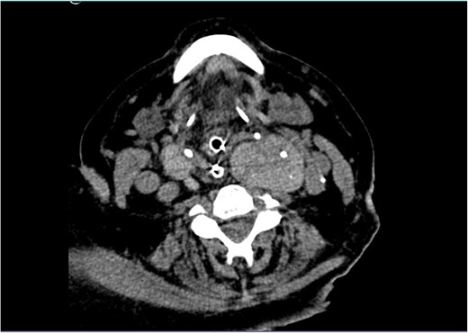
Neck CT showing a large goitre with extension to the left angle of the mandible.
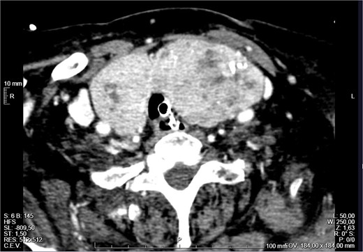
External compression of the thyroid and larynx by the mass with narrowing of its lumen.
The patient was admitted to the Intensive Care Unit (ICU). A positive Pemberton sign was found on physical examination (Fig. 3). The thyroid hormones were normal with a slightly decreased TSH. The case was discussed in a multidisciplinary meeting and surgical treatment was decided. On the fourth day of admission, the patient underwent a total thyroidectomy (Fig. 4), parathyroid autotransplantation, and tracheostomy. The procedure was uneventful. The histological examination revealed nodular hyperplasia of the thyroid with foci of micropapillary carcinoma (4 mm) in the right lobe. The patient was extubated and discharged from the ICU on the second postoperative day. She initiated a physical and speech therapy programme. The follow-up nasopharyngoscopies showed a slow but progressive improvement of the laryngeal oedema, which required a longer admission. After a few unsuccessful attempts, she was finally decannulated on the 39th postoperative day and was soon discharged home. She progressed well and two and a half years after the surgery she is asymptomatic, with no dysphonia or respiratory distress. She has a subclinical hyperthyroidism that has been monitored in annual follow-up appointments.
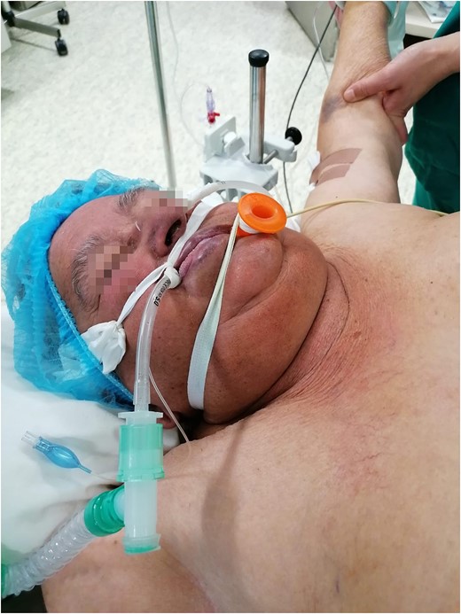
Positive Pemberton sign.
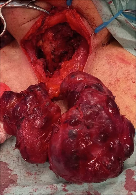
Surgical specimen after total thyroidectomy.
Discussion
AUAO caused by thyroid pathology usually occurs in association with anaplastic carcinoma [3–6], fast-growing tumours that can compromise the upper airway in early stages, either through external compression, direct invasion of the trachea or bilateral vocal cord paralysis [3, 4]. A benign multinodular goitre presenting with AUAO requiring urgent surgery, as presented here, is quite rare [2, 5, 6]. Its physiopathology is not yet fully understood, but it is believed to be multifactorial [2]. Haemorrhage inside the gland, upper airway infection or exacerbation of chronic respiratory diseases can be causes of respiratory distress in these patients [2, 3, 6].
AUAO can occur regardless of size, symptoms, length of thyroid disease, or a plunging goitre [7]. A neck CT is the gold standard for the diagnosis [5], considering the clinical presentation can mimic an exacerbation of the patient’s respiratory comorbidities [6]. There seems to be a lack of correlation between evidence of airway obstruction in pulmonary function tests and the narrowing of the trachea’s diameter in CT scans [7].
The management of this life-threatening condition is controversial [3, 6]. It is important to assess thyroid function prior to the surgery, even in the emergency setting [6]. However, the first step should be prompt intubation [3, 6], like it was done in this case. Medical treatment includes oxygen therapy, humidification, bronchodilators, corticosteroids, and adrenaline nebulization. In what concerns surgical therapy, urgent thyroidectomy is the gold standard procedure and its safety in specialized centres has already been shown [2, 5].
In the postoperative period, most patients will require mechanical ventilation for a few more days, while others with persistent laryngeal oedema and tracheomalacia might need an endotracheal stent or a tracheostomy [6]. In this case, the patient required almost 40 days of admission due to the laryngeal oedema that prevented an early removal of the tracheostomy. Tracheomalacia is a very rare condition where the trachea’s lumen is reduced to <50% due to prolonged compression of the airway by a large goitre, resulting in deterioration and weakening of the tracheal rings which makes the trachea more prone to collapse after thyroidectomy [8]. This can lead to postoperative AUAO and must be managed immediately. Despite not having occurred in the described case, another postoperative complication is the development of a cervical haematoma [9, 10]. For many years it was assumed that post-thyroidectomy haematoma caused AUAO through direct compression, but current literature states the main cause of airway compromise is the laryngeal oedema induced by obstruction of venous outflow [9, 10].
In conclusion, patients with large multinodular goitres are at risk of airway compromise, especially when they have other comorbidities. AUAO is a medical emergency and should be addressed immediately. As shown in the management of this case, the priority is securing the airway. Afterwards, medical treatment should be initiated followed by early thyroidectomy. Postoperative monitorization for laryngeal oedema and tracheomalacia is crucial and rehabilitation programs are essential to a full recovery and return to normal life.
Conflict of interest statement
None declared.
Funding
None declared.
