


Abstract
Gallbladder hemangiomas are extremely rare benign tumors. Eighteen cases have been reported in the literature, with only two associated with gallstones. We report the third case of a gallbladder hemangioma associated with gallstones in a 54-year-old Moroccan woman, along with a comprehensive review of the literature. Gallbladder hemangiomas are likely underdiagnosed, underscoring the need for careful examination of cholecystectomy specimens.
Introduction
Hemangiomas are benign vascular tumors primarily located in the skin. Gallbladder hemangiomas are exceptionally rare, with only 18 cases reported in the literature, including 2 associated with gallstones [1]. We present the third case of gallbladder hemangioma associated with gallstones in a 54-year-old Moroccan woman.
Case report
A 54-year-old Moroccan woman with no significant medical history presented with right upper quadrant pain lasting for two months, accompanied by nausea but no fever, weight loss, or other systemic or digestive symptoms. Abdominal ultrasonography revealed gallstones. The patient underwent laparoscopic cholecystectomy. Macroscopic examination revealed a gallbladder measuring 8.6 cm in length and 2.3 cm in width, with a slightly thickened wall and the presence of gallstones. Microscopic examination showed chronic diverticular cholecystitis and a vascular proliferation measuring 10 mm, composed of variably sized, often ectatic blood vessels containing red blood cells (Fig. 1). The vessels were lined by a single layer of regular endothelium (Fig. 2). The patient’s postoperative course was uneventful.
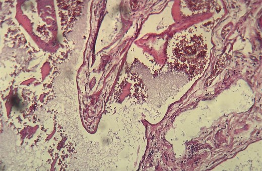
Histopathological examination showing ectatic blood vessels containing red blood cells.
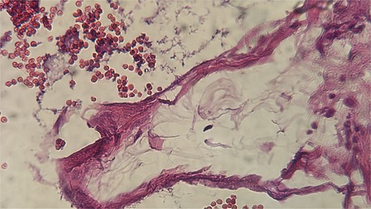
The vessels were lined by a single layer of regular endothelium.
Discussion
Hemangiomas are predominantly located in the skin, and gallbladder hemangiomas are exceedingly rare. We identified 18 cases in the literature, 2 of which were associated with gallstones (Tables 1 and 2), with our case being the third. Clinically, gallbladder hemangiomas are usually asymptomatic but can present with abdominal pain [1, 2], hemoperitoneum [17], or jaundice [18]. Abdominal ultrasonography, the initial imaging modality for hepatobiliary diseases, may fail to detect hemangiomas, particularly when gallstones are present, as seen in our case. Gallstones can explain the symptoms or pose a differential diagnostic challenge with neoplastic pathologies [18]. Macroscopically, gallbladder hemangiomas can be asymptomatic or appear as violaceous hemorrhagic tumors [1]. Microscopically, they resemble hemangiomas in other locations, showing vascular proliferation with ectatic vessels containing red blood cells and lined by regular endothelium. The histopathological diagnosis is generally straightforward, but in cases of endothelial detachment, immunohistochemistry using endothelial markers can aid diagnosis. The treatment is surgical, consisting of cholecystectomy. In cases with bleeding risk or suspected malignancy, laparotomy exploration is recommended [18]. Our case represents the third reported instance of gallbladder hemangioma associated with gallstones. Gallbladder hemangioma is an extremely rare benign tumor that is likely underdiagnosed. Careful macroscopic and microscopic examination of cholecystectomy specimens is essential for accurate diagnosis.
Acknowledgements
The authors acknowledge anyone who has participated in the care of this patient directly or indirectly.
Conflict of interest statement
None declared.
Funding
None declared.
Consent for publication
Written informed consent was obtained from the patient for publication of this case report and any accompanying images. A copy of the written consent is available for review by the Editor-in-Chief of this journal.
Guarantor
Imane Boujguenna is acting as a guarantor for this manuscript.
