-
PDF
- Split View
-
Views
-
Cite
Cite
Shivani Raju, Shravya Kashyap, Afnan W M Jobran, Shrivarshini K, Shravya N, A rare presentation of hematosalpinx with torsion in a thirteen year-old virgin: a case report, Journal of Surgical Case Reports, Volume 2024, Issue 9, September 2024, rjae549, https://doi.org/10.1093/jscr/rjae549
Close - Share Icon Share
Abstract
A medical condition called hematosalpinx causes an accumulation of blood within the fallopian tube. It is usually seen in patients with ectopic pregnancy. Inflammatory disease of the pelvis, tubal cancer, pelvic trauma, and endometriosis are further causes. Here, we report a unique case of hematosalpinx with associated tubal torsion in a 13-year-old female lacking any previously reported contributing causes. She is celibate and presented with abdominal pain and fever. Beta-hcg was not present, and her menstrual cycle was regular. Pelvic ultrasound sonography revealed a large cyst and was suggestive of a right paraovarian cyst. An exploratory laparotomy was performed and a giant hematosalpinx was observed in an otherwise normal ovary. In conclusion, although very rare in adolescence, hematosalpinx must be considered in the differential diagnosis. This unusual instance highlights new concerns regarding the pathogenesis of hematosalpinx.
Introduction
Hematosalpinx is the accumulation of blood in fallopian tubes. It is a rare condition that results from a distended blood-filled tubal lumen with a reported incidence of 1.08% to 1.8% [1]. Ectopic pregnancy is the main factor contributing to this condition; however, other factors include pelvic inflammatory disease (PID), endometriosis, and pelvic trauma [1, 2]. Abdominal pain, unusual vaginal bleeding, and irregular menstruation are just a few of the symptoms that might appear with hematosalpinx, which has a number of underlying etiologies. One or both fallopian tubes may be affected by the disorder, which can be unilateral or bilateral [3].
Isolated fallopian tube torsion is a rare cause of acute lower abdomen pain with a rare underlying etiology. This was a rare case of isolated right fallopian tube torsion with hematosalpinx. Following are some hypothesized causes of fallopian tube torsion: physiologic abnormalities, such as abnormal peristalsis or periovulatory spasm; hemodynamic abnormalities, such as long mesosalpinx, tortuous dilated tube (hydro- or hematosalpinx); tubal mass (tubal neoplasm); and adnexal mass (adjacent ovarian or paraovarian tumor), such as Sellheim hypothesis (sudnexal venous congestion); trauma; prior surgery; or illnesses including PID; pelvic adhesion; tubal ligation; gravidity; and an enlarged or uterus [4].
We report a case of hematosalpinx with bleeding in a 13-year-old girl who had an isolated right fallopian tube that was effectively treated with a partial salpingectomy.
Case presentation
A 13-year-old celibate girl was referred to our hospital after enduring 2 months of abdominal pain in the right lumbar and right iliac area. She explains the pain started gradually, was intermittent, and radiates to her right leg in a spasmodic manner. Before approaching the hospital, she also had related nausea, vomiting, and fever for 5 days. Two years ago, she reached menarche; albeit having a regular cycle, she experienced dysmenorrhea and clots. She did, however, have burning micturition and heavy bleeding during periods. Upon self-examination, a felt mass grew larger over time. There were no observed extragenital abnormalities or comorbidities.
Throughout the examination, she was conscious, cooperative, and well-oriented to the time, place, and person. There was pallor. The umbilicus was soft to the touch, centered in the abdomen, and free of any scars or pulsations. There was pain and a suprapubic mass in the lower right quadrant of the abdomen. Upon assessment, no further complications were found.
An additional examination of the electrocardiogram showed sinus tachycardia. The ultrasound of the right ovary showed a large, clearly defined anechoic cystic lesion measuring 10.9 × 10.00 × 11.1 cm (volume: 641 cc), with only a few eccentric cystic lesions and no internal separation or debris. The right fallopian tube is edematous, deformed, and filled with free fluid medially to the previously noted lesion. A pre-op diagnosis of paraovarian cyst was affirmed and an exploratory laparotomy was proposed.
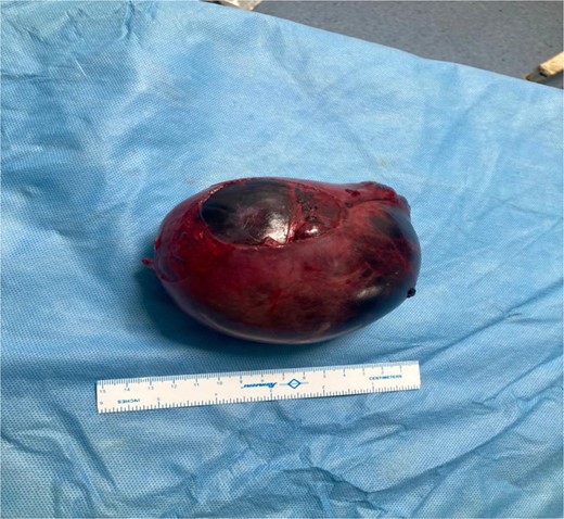
Intraoperatively, on the right side, a 13 × 12 × 8 cm hematosalpinx—confirming as the post-op diagnosis—with a total of three coils of torsion was found; it was detorted, clamped, cut, and ligated, completing a partial salpingectomy. On histopathological examination, the gross features showed a single soft tissue mass that was gray–black in color. The cut section exhibits brown fluid and is uniloculated, whereas the external surface was dilated. Microscopic characteristics revealed a fallopian tube with a hemorrhagic, dilated lumen. The mucosa presents little plicae and is bordered by low cuboidal to columnar epithelium, while the wall displays muscular thinning and hemorrhage.
The surgery was uneventful (Figs 1 and 2) and there were no postoperative complications and infections; the patient progressed toward recovery.


Discussion
Hematosalpinx is the accumulation of blood in the distended tubal lumen, which is a rare case by itself. Hematosalpinx accompanied with tubal torsion in a young girl makes it even more complex. The average incidence of isolated tubal torsion is 1 in 1500 000 women, therefore being a rare occurrence. It can be noticed during an array of reproductive periods, including youth, pregnancy, and the perimenopausal and menopausal stages [1, 4].
The typical etiologies in non-pregnant women are tubal endometriosis, ongoing salpingitis, torsion, and tumors. Non-tubal congenital and acquired obstructive causes include tumors, ablation, adhesions, imperforate hymen, and uterine abnormalities [5–7]. It is essential to distinguish both intrinsic and extrinsic predisposing variables in the etiology of fallopian tube torsion. A tubal tumor, hydro-, pyo-, or hematosalpinx, an exceedingly long tube, ovulatory or premenstrual congestion, and prior sterilization are examples of the former. Ovarian tumors, uterine enlargement resulting from pregnancy or fibroids, adhesion, and trauma are a few such extrinsic factors [4, 8].
Tubal torsion can occur in the normal or the diseased lumen, mostly commonly occurring in the latter [9]. Hematosalpinx may have no visible signs or present pelvic pain which may be accompanied by vaginal bleeding or not. Hemoperitoneum and tubal rupture akin to an acute abdomen [10]. In our case, the patient presented with acute lower abdominal pain, with no vaginal bleeding. On examination, an abnormal mass was palpable over the abdomen which was not ruptured.
Accurate diagnosis of the condition is crucial as it can be overlooked to other differentials, such as tubal ectopic pregnancy, endometriosis, tubal carcinoma, retrograde menstruation, or uterine cervical stenosis. Ultrasound, computed tomography (CT) and magnetic resonance imaging are the diagnostic tests performed to confirm the diagnosis. A dilated fallopian tube may be observed on ultrasound, and altered blood products within the tube are frequently observed as homogeneous low-level echoes. Yet, during a CT scan, blood products within a dilated fallopian tube may appear as high attenuation [11]. Along with the radiological diagnostics, laparoscopy is performed, which is considered a gold standard. The young female was diagnosed with a differential of paraovarian cyst, and was first suggested with exploratory laparotomy, by which a precise diagnosis can be confirmed during the surgery.
The initial course of action in managing hematosalpinx is to surgically eliminate the diseased fallopian tube (salpingectomy), or if isolating the ovary is not possible, to remove the tubovarial formation. If the formation ruptures, the abdominal cavity is then cleaned. Access by laparoscopy is desirable. The outlook for survival is good with prompt surgical intervention. Salpingectomy improves the success rate of assisted reproductive technologies, which helps the desired conception to start [12]. Anti-inflammatory (NSAIDs, dexamethasone) and enzyme resorption (hyaluronidase) therapy has been suggested during the postoperative period.
Conclusion
The careful management of pelvic organ inflammatory conditions, the practice of personal and intimate cleanliness, and routine preventative exams are all important to prevent the development of hematosalpinx. It is always suggested for regular gynecological visits from an early age to treat and cure any abnormalities to prevent the further development that could hamper with the well-being of an individual.
Conflict of interest statement
None declared.
Funding
None declared.
References
- pregnancy
- ultrasonography
- abdominal pain
- cancer
- ectopic pregnancy
- adolescent
- endometriosis
- fever
- chorionic gonadotropin, beta subunit, human
- cysts
- differential diagnosis
- fallopian tubes
- menstrual cycle
- sexual abstinence
- ovary
- pelvis
- inflammatory disorders
- pelvic ultrasonography
- pelvic injuries
- laparotomy, exploratory
- paraovarian cyst


