-
PDF
- Split View
-
Views
-
Cite
Cite
Katie Nightingale, Emily Clough, Paul Goldsmith, Joshua Richard Burke, Peritoneal inclusion cyst presenting as an umbilical hernia: case report and systematic review of the literature, Journal of Surgical Case Reports, Volume 2024, Issue 5, May 2024, rjae258, https://doi.org/10.1093/jscr/rjae258
Close - Share Icon Share
Abstract
Peritoneal inclusion cysts (PICs) are a rare and benign condition of uncertain pathogenesis. The fluid-filled, mesothelial-lined cysts manifest within the abdominopelvic cavity. This case report details an unusual occurrence of a 97 mm PIC- presenting as an umbilical hernia- in a 26-year-old male patient with no prior surgical history. Following pre-operative cross-sectional imaging, this was managed through open excision without complication. A systematic review of the literature highlighted 30 previous cases [26F, 4M] with a mean age of 34 years (std ±15.4) and a median diameter of 93 mm [IQR, 109 mm]. A total of 53% (n = 16) of cases had a history of previous abdominal surgery. Surgical excision is safe and laparoscopic modality should be considered (<1% recurrence). Accepting the limited evidence base, image guided drainage should be avoided (50% recurrence, n = 2).
Introduction
Peritoneal inclusion cysts (PICs), also known as multicystic peritoneal mesotheliomas, are a rare and benign condition of uncertain pathogenesis [1]. The fluid-filled, mesothelial lined cysts manifest within the abdominopelvic cavity and can vary in size. In the few case reports presented, they are predominantly observed in women of childbearing age and have been associated with chronic peritoneal inflammation stemming from a variety of aetiologies and prior surgical interventions [2, 3]. To date, surgical excision has been the only considered definitive treatment [4]. This paper details a case of PIC presenting as an umbilical hernia, reported as per the for CAse REports (CARE) guidelines. [5], and a systematic review of the literature compliant with Preferred Reporting Items for Systematic Review Meta-Analysis (PRISMA) guidelines [6].
Case report
We present the case of a 26-year-old man who presented to the general surgery outpatient clinic with intermittent peri-umbilical pain, and the presence of a soft swelling palpable at his umbilicus. There was a positive cough impulse consistent with a true umbilical hernia and the patient described no previous medical or surgical history. An abdominal ultrasound scan visualized a large thin-walled serous fluid collection tracking into the peritoneal cavity (95 mm × 87 mm × 97 mm) (Fig. 1). Computed tomography (CT) of the abdomen and pelvis with contrast reported a large bi-lobed cystic mass centred at the right side of the mesentery, with herniation of part of the cyst along the umbilicus. Displacement of the small bowel with anterior extension into the abdominal wall was seen with the suggestion of posterior extension into the right retroperitoneal space (Figs 2 and 3). The patient underwent routine pre-operative work up and the cyst was excised through a midline para-umbilical laparotomy (Fig. 4) given the concern of retroperitoneal involvement. Intra-operatively, the hernia neck and the root of the cyst were found at the base of the umbilical cicatrix with no attachment to the mesentery. The cyst was loculated and filled with clear fluid. It was dissected off the peritoneal tissues and off from the posterior umbilical skin prior to removal. There was evidence of rupture of one of the locules and clear fluid was drained.
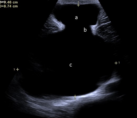
Ultrasound image demonstrating fluid collection within the umbilicus (a) tracking into abdominal cavity (c) originating from the cicatrix (b).
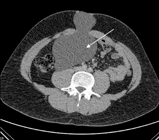
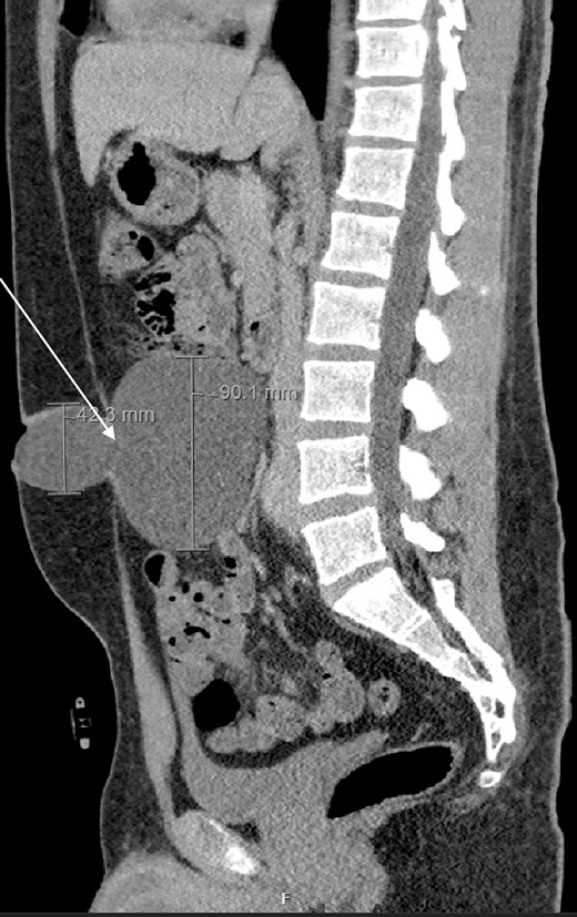
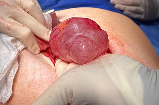
PIC was excised through midline laparotomy given the suggestion of retroperitoneal involvement on pre-operative imaging.
Histological examination demonstrated a paucicellular fibrous wall including frequent small blood vessels. The outer surface constitutionally comprised of loose fibrous tissue mixed with adipose tissue, and the lining was composed of a monolayer and- focally- a double layer of mesothelial cells. This showed no significant atypia, and a diagnosis of PIC with no evidence of neoplasia was made. He was discharged without complication, with a follow up CT planned at 6 months.
Systematic search
The MEDLINE, EMBASE electronic databases published between 1 January 1947 and 1January 2024 were searched using the term ‘peritoneal inclusion cyst’. All studies which reported the case of a PIC were considered. KN independently retrieved article abstracts with JB cross-checking. The full texts of potentially eligible studies were retrieved and independently assessed for eligibility by KN and JB according to predefined inclusion criteria. Any disagreement was resolved through discussion with PG. Initial search yielded 61 full text articles from 1946 to 2023 for review as displayed in the PRISMA Flow Chart (Fig. 5), with 30 included in the final analysis (Table 1).
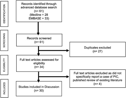
Preferred Reporting Items for Systematic Review Meta-Analysis (PRISMA) flow chart showing selection of reports for discussion.
| Author(s) . | Country . | Sex . | Age . | Max.Diameter (mm) . | Imaging . | Origin . | Intervention . | Complications . | Previous surgery . |
|---|---|---|---|---|---|---|---|---|---|
| Coulibaly et al. 2023 [7] | France | Female | 29 | - | PET | Pelvic cavity | Laparoscopic | None | Not documented |
| AlTamimi et al. 2023 [3] | United States | Female | 18 | 150 | CT | Ovary | Open | Failed IR drainage | Previous open bladder repair |
| Guo et al. 2023 [8] | England | Female | 43 | 73 | USS | Vaginal/uterine space | Open | None | Previous fibroid and ovarian cyst removal. Previous appendicectomy |
| Ivanova et al. 2022 [9] | Russian Federation | Female | - | - | - | Ovary | Laparoscopic | - | Not documented |
| Subramonian et al. 2021 [10] | Netherlands | Female | 14 | 87 | USS | Recto-vaginal space | Laparoscopic | None | Previous laparotomy (ileal perforation) |
| Cosgrove et al. 2021 [11] | Northern Ireland | Male | 41 | 86 | CT/MRI | Mesentery to the sigmoid colon | Open | None | Not documented |
| Katebi et al. 2021 [12] | Netherlands | Female | 44 | 200 | CT | Recto-vaginal space | Laparoscopic | None | Not documented |
| Kato et al. 2010 [13] | Japan | Female | 21 | - | Not recorded | Pelvic cavity | Not documented | Not recorded | Not documented |
| Wolf et al. 2020 [14] | Netherlands | Female | 72 | - | CT/MRI | Pelvic cavity | Open | Recurrence | Previous hysterectomy |
| Napoe and Rardin 2020 [15] | England | Female | 47 | 50 | CT | Uterine | Open | None | Hysterectomy |
| Katebi et al. 2019 [12] | Netherlands | Female | 44 | 200 | MRI | Recto-vaginal space | Laparoscopic | Not recorded | Not documented |
| Pereira 2019 [16] | England | Female | 34 | 107 | MRI | Pelvic cavity | Spontaneously ruptured | None | Previous robotic assisted endometrial resection, appendicectomy and adhesiolysis |
| Mishra et al. 2018 [17] | India | Male | 43 | 210 | USS/CT | Mesocolon | Laparoscopic | None | None |
| De Luca et al. 2017 [18] | Netherlands | Male | 49 | 80 | USS | Mesenteric root / precaval | Laparoscopic | None | Previous open appendicectomy |
| Sato et al. 2017 [19] | Ireland | Female | - | - | - | Ovary | Laparoscopic | - | 2 prior laparotomies |
| Singh et al. 2015 [2] | India | Female | - | - | CT | Ovary | Open | None | Previous bilateral tubal ligation |
| Trehan. 2014 [20] | England | Female | 51 | 57 | MRI | Recto-vaginal space | Laparoscopic | None | Previous bilateral tubal ligation |
| Kanasugi et al. 2013 [21] | Japan | Female | 31 | - | MRI/USS | Pelvic cavity | Open | None | Not documented |
| Saxena et al. 2011 [22] | United States | Female | 7 | 80 | USS/MRI | Ascending colon mesentery | Laparoscopic | None | Not documented |
| Ho-Fung et al. 2011 [23] | United States | Female | - | - | Pelvic cavity | Cystocentesis | None | Not documented | |
| Dillman and DiPietro 2009 [24] | Germany | Female | 16 | - | USS | Ovary | Laparoscopic | None | Multiple previous adhesiolysis |
| Vallerie et al. 2008 [25] | United States | Female | 29 | 217 | USS | Pelvic cavity | Cystocentesis | Recurrence | 2 previous exploratory laparotomies |
| Advincula and Hernandez 2006 [26] | United States | Female | 36 | 100 | CT | Recto-vaginal space | Laparoscopic | None | Previous hysterectomy and exploratory laparotomy |
| Phupong et al. 2005 [27] | Germany | Female | 33 | 61 | USS | Ovary | Laparoscopic | None | Not documented |
| Nayak et al. 2005 [28] | India | Female | 26 | 0 | Found during caesarean | Pelvic cavity | Open | None | Previous caesarean section |
| Durak et al. 2005 [29] | United States | Male | 32 | 140 | CT | Pelvic cavity | Open | Not documented | |
| Toprak et al. 2004 [30] | United States | Female | 24 | - | USS/CT/MRI | Adnexa | Open | None | None |
| Omeroglu and Husain. 2001 [31] | United States | Female | 31 | 75 | CT | Ovarian | Open | None | Previous myomectomy |
| Brustmann 2000 [32] | United States | Female | 21 | 200 | - | Mesentery of terminal ileum | Open | None | Not documented |
| Lamovec and Sinkovec 1996 [33] | England | Female | 68 | 200 | CT/MRI | Mesentry of descending colon | Open | None | Not documented |
Discussion
This is the first documented case of a PIC originating from the umbilical cicatrix. Thirty reported cases of PIC have been identified, 26 female and 4 male, with a mean age at presentation of 34 years (std ±15.4) and a median diameter of 93 mm [IQR, 109 mm]. A total of 53% (n = 16) of cases had a history of a previous abdominal surgery. Thirteen were excised via laparotomy, 13 were excised laparoscopically, 2 were drained under Interventional radiology guidance, 1 ruptured spontaneously with no further sequalae, and 1 case did not report their operative approach. Of the two cases that were drained, one recurred. Of those excised, only one recurred (median follow up 24 months (range 0–60 months)).
The most common organ of origin described was ovarian- in 23% of cases (N = 7), followed by the recto-vaginal space (17%, n = 5). In seven cases (23%) the origin was broadly defined as the pelvic cavity. Other origins included mesentery of small and large bowel, adnexa, and the vaginal-uterine space.
This case underscores the atypical presentation of a PIC in a surgically naïve male patient, emphasisizing the importance of pre-operative imaging in those presenting with atypical features of hernia; this instance, skin changes and discharge in the absence of previous abdominal surgery. Existing literature supports surgical excision rather than drainage given the risk of recurrence. Surveillance and required follow up specificities are currently unknown and further research is needed to determine this.
Author contributions
KN completed the data collection, statistical analysis of literature and drafting of manuscript. EC, PG & JB contributed to case conception, and manuscript editing.
Conflict of interest statement
The authors declare no conflicts of interest.
Funding
None declared.
Consent
Informed patient consent was obtained and no identifiable details are mentioned in this report.


