-
PDF
- Split View
-
Views
-
Cite
Cite
Akio Sakamoto, Takashi Noguchi, Shuichi Matsuda, Extending the usefulness of the Stryker Growing Prosthesis in pediatric patients, Journal of Surgical Case Reports, Volume 2024, Issue 2, February 2024, rjae066, https://doi.org/10.1093/jscr/rjae066
Close - Share Icon Share
Abstract
Osteosarcoma is a highly invasive primary bone tumor that predominantly occurs in childhood and adolescence. The Stryker Growing Prosthesis provides a means of reconstructing large bone defects resulting from bone resection in skeletally immature patients. This device can be expanded as the patient grows. The possible length of extension depends on the length of the prosthesis. Because further expansion was not possible, by turning the adjustable part of the extension back to zero and adding a new permanent extension allow the prosthesis to be further adjusted as growth ensues. Using this method/device only, a separate endoprosthesis was required to be attached onto the extension. Therefore, the applicable cases are limited, because of the fact that extensive resection usually means total femoral replacement is best indicated. However, this method is still useful for reducing the number of revision surgeries in such cases. This reduces costs and increases savings for insurers/countries.
Introduction
Osteosarcoma is a highly invasive primary bone tumor that predominantly occurs in childhood and adolescence. It is a highly aggressive malignancy that usually metastasizes to the lungs [1]. A regimen consisting of cisplatin, doxorubicin, and high-dose methotrexate can result in long-term disease-free survival rates of ~70% [2].
In the case of limb-sparing surgery, a prosthesis can be used for reconstructive purposes after sarcoma resections of the extremities because it provides mechanical stability and postoperative rehabilitation [3]. Osteosarcomas typically occur at the metaphysis, adjacent to the growth plates of bones, the latter of which are responsible for bone lengthening. Resection at the tumor site following the fitting of a prosthesis tends to lead to a leg-length discrepancy, particularly in young children [3].
The Stryker Growing Prosthesis provides a means of reconstructing large bone defects resulting from bone resection in skeletally immature patients. This device can be expanded as the patient grows. The system consists of articular components (proximal femur, distal femur, and/or proximal tibia), extension components, and stems. Modular implants can also be combined. The prosthesis is designed before the operation, based on the amount of bone to be resected and an estimation of the remaining growth of the patient. It is possible to expand this prosthesis using a minimally invasive procedure; however, the maximum extension depends on the length of the prosthesis.
Case report
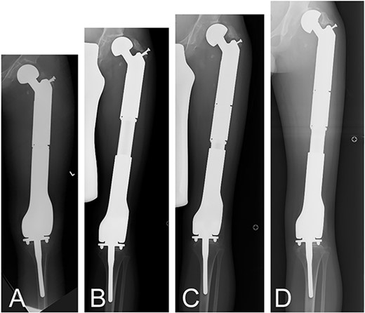
A 5-year-old boy presented with osteosarcoma of the left distal femur. After preoperative chemotherapy, a wide excision was performed at our institution. The bone containing the tumor cells was frozen using liquid nitrogen, resected, reused, and a plate was fixed [4, 5]. The reused bone was fractured. Ultimately, the entire femur was replaced with an endoprosthesis when the patient was 8 years old. For this case, the proximal femoral component was 120 mm in length and was not an expandable prosthesis. The distal femoral component was 160-mm long and the expandable prosthesis had a maximum expandable length of 62 mm (Fig. 1A). A total dilation of 56 mm was achieved with three surgeries at the age of 11 years (each expansion was 20, 20, and 14 mm) (Fig. 1B). Because further expansion was not possible, by turning the adjustable part of the extension back to zero and adding a new permanent 60-mm extension, allows the Stryker Growing Prosthesis to be further adjusted as growth ensues at the age of 12 years. The adjustable section of the prosthesis then had a maximum re-expandable length of 62 mm, and was expanded by an additional 14 mm at the operation (Fig. 1C). The total expansion was 62 mm over the course of five surgeries (each expansion; 15, 15, 10, 11, and 11 mm), resulting in a total of 122 mm of expansion from the time of initial total femoral replacement by the age of 13 years (Fig. 1D).

A 5-year-old boy presented with osteosarcoma of the left distal femur. After the tumor was resected, the defect was reconstructed by a recycled frozen autograft. The recycled bone was fractured, and total femoral replacement with the Stryker Growth Prosthesis was required when he was 8 years old (A). By 11 years of age his femur was extended by 80 mm (B). The extension was reduced to 0 mm, and 60-mm spacer was added (C), and then the Stryker Growth Prosthesis was further extended, resulting in a total of 122 mm of expansion from the time of initial total femoral replacement by the age of 13 years (D).
Discussion
The possible length of extension depends on the length of the prosthesis, and/or the length of the resection. Also, the maximum length of an extension prosthesis is longer for the integrated type with a stem than for the separate type “without a stem.” The expandable length of the proximal femur and the distal femur is determined by subtracting from the length of the resection 98 mm for the integral type and 158 mm for the separate type. Surgeons tend to use one whole prosthesis to obtain longer extensions.
Using this method/device only, a separate endoprosthesis was required to be attached onto the extension. Therefore, the applicable cases are limited, because of the fact that extensive resection usually means total femoral replacement is best indicated. However, this method is still useful for reducing the number of revision surgeries in such cases. This reduces costs and increases savings for insurers/countries.
Conflict of interest statement
None declared.
Funding
None declared.


