-
PDF
- Split View
-
Views
-
Cite
Cite
Amir H Sohail, Koral Cohen, Kimberly Ho, Sawyer Cimaroli, Collin E M Brathwaite, Patrick Shin, Incidental aortocaval fistula in the setting of an unruptured abdominal aortic aneurysm, Journal of Surgical Case Reports, Volume 2023, Issue 7, July 2023, rjad384, https://doi.org/10.1093/jscr/rjad384
Close - Share Icon Share
Abstract
An aortocaval fistula, a rare abnormal vascular communication between the aorta and inferior vena cava, is most commonly associated with abdominal aortic aneurysms (AAAs). Other factors leading to aortocaval fistula formation include atherosclerosis, collagen vascular diseases, vasculitis, hematogenous infections, prior spinal surgery, malignancy and radiation exposure. In rare instances, aortocaval fistulas may be discovered incidentally on abdominal imaging. We report an unusual case of an incidental aortocaval fistula in a 93-year-old male patient with an unruptured AAA, presenting with shortness of breath, malaise and lethargy. The patient had no other obvious risk factors for aortocaval fistula formation. Multidetector computed tomography angiography helped identify the fistula, and the patient was eventually transferred to hospice for comfort measures. This case highlights the importance of detailed imaging and preoperative planning in managing aortocaval fistulas and associated AAAs.
INTRODUCTION
An aortocaval fistula is an abnormal vascular communication between the aorta [generally an abdominal aortic aneurysm (AAA)] and the inferior vena cava (IVC) [1]. It is a rare entity, most commonly seen in the setting of a ruptured abdominal aorta aneurysms, with an estimated incidence ranging from <1% to as high as 10% [2–5].
Other factors associated with aortocaval fistula formation include atherosclerosis, collagen vascular disease (i.e. Marfan, Ehler–Danlos syndrome), vasculitis (e.g. polyarteritis nodosa), hematogenous infections, prior spinal surgery, malignancy and radiation exposure [2, 6]. Penetrating trauma or spinal injury with thoracolumbar translation may also lead to aortocaval fistula formation. Rarely, IVC dissection or IVC filters have been reported to result in an aortocaval fistula [2].
In extremely rare instances, aortocaval fistulas may be seen as an asymptomatic incidental aortocaval finding on abdominal imaging. We report a rare case of an incidental aortocaval fistula in a patient with an unruptured AAA, and no other obvious inciting factor.
CASE DESCRIPTION
A 93-year-old male with a medical history of asthma and hypertension presented with complaints of shortness of breath, malaise and lethargy. Social history was significant for smoking. Review of symptoms was otherwise unremarkable.
Upon initial evaluation, vital signs were significant for bradycardia, hypotension and tachypnea. Physical examination revealed an ill-appearing, tachypenic male, using accessory muscles to breathe. The abdomen was soft, nontender and nondistended, without guarding or rebound tenderness. A pulsatile, nontender epigastric mass was palpable. Pedal edema was noted.
Laboratory investigations were notable for transaminitis (aspartate aminotransferase 1152 IU/L, and alanine transaminase 434 IU/L), elevated lactate dehydrogenase (2642 units/L) and elevated serum lactic acid (9.6 mmol/L), which uptrended despite fluid resuscitation. Serum creatinine was elevated. A rapid polymerase chain reaction test for SARS coronavirus was positive. A transthoracic echocardiogram was performed and showed a normal left ventricular ejection fraction (70%). The left ventricular filling pattern was pseudonormal.
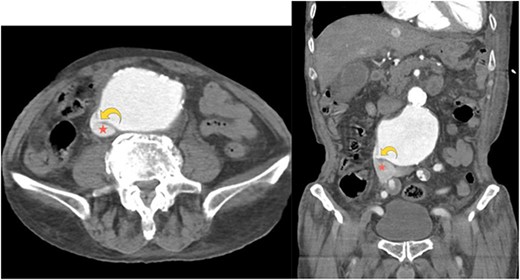
A right-upper quadrant abdominal ultrasound demonstrated an AAA, measuring 5.5 cm × 8.5 cm. Computed tomography angiography of the abdomen and pelvis showed a 9.2 cm aneurysm of the infrarenal abdominal aorta compressing the IVC (Fig. 1a). Furthermore, an aortocaval fistula was noted (Fig. 1a and b). There was no evidence of an aortic rupture or dissection. Of note, the dimensions and anatomy of AAA were amenable to endovascular repair.

Axial computed tomography angiography image showing a small focal defect in the aortic wall (curved yellow arrow). Abnormal early opacification of the IVC (red star) can be noticed in this arterial phase study. These findings are consistent with an aortocaval fistula. No retroperitoneal hematoma is observed.
The patient was placed on bilevel positive airway pressure respiratory support and required vasopressors due to hemodynamic instability. He went into cardiopulmonary arrest, and the return of spontaneous circulation was rapidly achieved. A family meeting was held and a decision was made to transfer the patient to hospice for comfort measures. The patient later expired.
DISCUSSION
The pathogenesis of aortocaval fistula involves degeneration and pressure necrosis of the aortic wall, most commonly in the setting of an AAA [2, 7]. This increases inflammation of the IVC and aortic vessel walls resulting in adherence of the AAA to the IVC. Eventually, the effacing walls erode and generate a fistula between the aorta and the IVC [1]. As such, the aortocaval fistula is a site of weakness, and over 80% of aortocaval fistulas are related to ruptured AAA [8]. Although uncommon, an AAA can rupture into the IVC in addition to the retroperitoneum, gastrointestinal tract and intraperitoneal cavity [9].
As noted above, very few aortocaval fistulas are incidentally discovered on cross-sectional abdominal imaging in association with an unruptured AAA and without another inciting factors [10]. Our extensive literature search revealed only one previously reported similar case of an aortocaval fistula associated with an unruptured AAA [10]. Interestingly, our patient did not have any of the other risk factors associated with aortocaval fistula formation, including severe atherosclerosis, prior surgical interventions, trauma or connective tissue disorders, etc. Furthermore, in our patient, imaging did not reveal a large amount of calcification near the aortocaval fistula.
Signs and symptoms of aortocaval fistula vary based on its chronicity. In the rare incidence of a AAA rupture into the IVC, complications such as high output cardiac failure, renal insufficiency, deep vein thrombosis, pulmonary embolism and shock may result [7, 8]. Refractory right-sided heart failure, abdominal bruit, pulsatile abdominal mass, back pain, hematuria/oliguria, wide pulse pressure and regional venous hypertension particularly in the setting of a known AAA or recent AAA repair should raise suspicion for aortocaval fistula [11]. Rarely, venous hypertension resulting from aortocaval fistula may manifest as lower extremity edema, which was seen in our patient.
Aortocaval fistulas are best identified on multidetector computed tomography angiography. Presence of contrast in both the aorta and IVC (on arterial phase images), which was seen in our patient, is pathognomonic for arteriovenous fistula. However, in case of compression of IVC from an AAA and no obvious fistulous tract, presence of contrast in iliac or renal veins on arterial phase and loss of aortocaval fat plane may be indicative of an aortocaval fistula [12].
Repair of aortocaval fistula and the associated AAA may be performed endovascularly (aortic stent graft, IVC stent graft or occluder plug) or via open surgery. This would have been the preferred approach in this case if the patient’s acute pathology and respiratory failure had resolved; his presentation was unrelated to the AAA and the aortocaval fistula. A 2022 meta-analysis revealed that of 110 cases of aortocaval fistula secondary to AAA rupture, 78% of cases were treated with an open approach, while the remaining 22% were treated endovascularly. Although 30-day survival was higher in the patients that were treated with an endovascular approach (97.6 vs. 87.5%), the reintervention rate was higher in those treated minimally invasively (35.7 vs. 2.5%) [13]. Data suggest that in case of open surgical repair, preoperative identification of aortocaval fistula is associated with a significantly lower mortality rate, highlighting the importance of preoperative planning and detailed imaging [14].
CONFLICT OF INTEREST STATEMENT
The authors declare no conflicts of interest.
FUNDING
No funding was received for this study.
ETHICAL APPROVAL
Patient consent was obtained, and all identifying information has been removed to ensure confidentiality.
References
- abdominal aortic aneurysm
- angiogram
- aorta
- atherosclerosis
- multidetector computed tomography
- dyspnea
- collagen-vascular diseases
- vasculitis
- cancer
- radiation exposure
- fatigue
- pathologic fistula
- hospice
- preoperative care
- inferior vena cava
- infections
- diagnostic imaging
- lethargy
- abdominal imaging
- spinal procedure
- comfort measures
- aortocaval fistula


