-
PDF
- Split View
-
Views
-
Cite
Cite
Saleh A Nedjim, Youssef Bencherki, Abdellah Nachid, Mehdi Safieddine, Mouad ElBadr, Oussama Moumen, Amine Moataz, Mohamed Dakir, Adil Debbagh, Rachid Aboutaieb, Hematuria complicated by urinary retention revealing post-radiotherapy gangrenous cystitis with peritoneal involvement: an exceptional case in current urological practice, Journal of Surgical Case Reports, Volume 2023, Issue 7, July 2023, rjad379, https://doi.org/10.1093/jscr/rjad379
Close - Share Icon Share
Abstract
Gangrenous cystitis is a pathology that is rarely encountered in current urological practice. It is due to necrosis of the bladder wall, which may be superficial or involve the entire wall. Its exact pathogenesis is unknown, but several factors make its diagnosis based on cystoscopy or imaging. Its surgical treatment depends on the operative finding. In this report, the authors report a case of gangrenous cystitis in a 73-year-old patient with a history of prostatectomy and radiotherapy whose main manifestation was urinary retention. The diagnosis was made by computed tomography scan. Surgical exploration confirmed the diagnosis, thus imposing a partial cystectomy with an omentum base plasty. Despite a good postoperative clinical and biological evolution, the patient died in the intensive care unit following respiratory distress and ventricular tachycardia. This case reminds once again the high mortality associated with this pathology.
INTRODUCTION
Gangrenous cystitis is a state of necrosis that can affect one layer or the entire thickness of the bladder wall. It can be localized or extensive. If the mucosa, submucosa and muscularis are involved, it can lead to spontaneous rupture and acute peritonitis [1]. Rapid diagnosis is difficult because the symptomatology is not specific [1] and relies on imaging studies and cystoscopy [2]. In the majority of cases described, treatment consisted of partial or total cystectomy [3]. The mortality associated with this entity is high, reaching up to 35% [4]. In this report, the authors report a case of gangrenous cystitis in a 73-year-old patient with a history of prostatectomy and radiotherapy whose main manifestation was urinary retention. The diagnosis was made by computed tomography (CT) scan. Surgical exploration confirmed the diagnosis, thus imposing a partial cystectomy with an omentum base plasty. Despite a good postoperative clinical and biological evolution, the patient died in the intensive care unit following respiratory distress and ventricular tachycardia.
CASE PRESENTATION
This is a 73-year-old patient who underwent prostatectomy for prostatic adenocarcinoma 5 years ago with a Gleason score of 8 (4 + 4) and an initial protein specific antigen of 17 ng/ml. A total of 2 years after the surgery, he underwent adjuvant radiotherapy combined with hormone therapy. All of the above management was performed in another center. He presented to our center with acute urine retention and abdominal pain. Three days before admission, he reported the notion of hematuria with dysuria and transit disorders due to a matter arrest. The whole evolving in a context of decline of general state made of anorexia and asthenia. On admission, the patient was conscious but asthenic (performance status at 2), diffuse abdominal sensitivity and a painful bladder globe. The emergency treatment was a bladder catheterization with 100 cc of thematic urine with clots, a venous line with administration and a biological check-up (creatinine at 149 with K at 6.9 without electrical signs on the electrocardiogramm, blood cells at 14 000/mm3 and C reactiv protein (CRP) at 450).
The CT scan showed a full bladder with multiple dense, declining formations, with air bubbles in their breasts, not enhanced after injection of contrast medium. It is also the site of a dense, heterogeneous, non-decreasing formation at the level of its dome, with multiple air bubbles within it, protruding at the level of the peritoneal fat opposite the dome with parietal defect. Presence of a subperitoneal effusion with hydroaerobic levels in the bladder. Presence of a pneumoperitoneum of medium abundance with diffuse infiltration of the mesenteric fat (Fig. 1). The decision to refer to the operating room (OR) and explore was made.

CT images showing a deformed bladder with the presence of a hematoma and air bubbles and from the contents to the dome lateralized to the left. Pneumoperitoneum with peritoneal effusion.
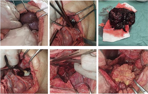
Initially, intraperitoneal exploration showed a discreet dilatation of the bowels without signs of suffering or obstruction. A minimal effusion was present. Subperitoneally, a hematoma was noted through the affected peritoneum (presence of a breach). In a second step, after opening the peritoneum covering the bladder, we found the presence of a hematoma and liquid. The first step was to aspirate the fluid and remove the hematoma. Discovery of a bladder breach of ⁓5 cm at the level of the dome lateralized to the left. The edges were necrotic and the wall very inflamed (Fig. 2). Extraction of the entire intravesical hematoma revealing the balloon of the bladder catheter. The edges are revived followed by cystorraphy and epiploplasty. Placement of a Redon drain, peritoneal closure and various walls.

Set of images showing the different operations from the demonstration of the hematoma through the peritoneum to the placement of the omentum after cystorraphy.
In the immediate postoperative period, the patient benefited from a renal purification session and was then sent to the surgical unit. The evolution was favorable from D1: the urine began to clear with a diuresis of 2400 cc on average per day, Redon to 30 cc. The evolution of biological parameters was favorable (creatinine at 83 with K at 5.1, blood cells at 11 700/mm3 and CRP at 324). On day 2, the patient was extubated with recovery of consciousness, apyretic, diuresis at 2 L and removal of the redon. Abruptly, he presented a disorder of consciousness with respiratory distress and ventricular tachycardia followed by cardiorespiratory arrest. Despite resuscitation measures, he could not be recovered and death was announced.
DISCUSSION
The first case of gangrenous cystitis was described by Willis in 1650 [5]. In the era of antibiotics, it has become an extremely rare entity [6]. As of 3 May 2023, entering the term gangrenous cystitis in English on Pubmed, only 67 results are displayed in the period between 1948 and 2023. This shows the rarity of this pathology.
The exact pathogenesis of bladder wall necrosis remains unknown. However, direct and indirect factors have been identified. Indirect factors are responsible for reduced blood flow and nutritional supply to the bladder, (chronic retention with overdistension, extravesical pressure and occlusion of major vessels). Direct factors are those leading to cell death due to damage to the bladder wall (chemicals injected into the bladder, pelvic irradiation and infection). Oniwa et al. reported a case of gangrenous cystitis 10 years after radiotherapy [7]. In our present case, indirect and direct factors were identified: retention, hematoma and radiotherapy. However, the radiotherapy was ˂10 years old.
The symptomatology of gangrenous cystitis is not specific. Suprapubic pain, frequency and dysuria, lower abdominal discomfort, pain directed toward the distal urethra, microscopic or macroscopic hematuria, pyuria or urosepsis are differential diagnoses for a variety of conditions, thus delaying rapid diagnosis [1].
Imaging studies (ultrasound, CT, cystography) and cystoscopy can assist in the diagnosis [3]. Imaging performed on an emergency basis allowed the diagnosis of a bladder breach. Despite the performance of cystography, there was no extravasation of the contrast medium. This could be explained by the sealing of the breach by the hematoma.
In our case, the involvement was extra trigonal. In the vast majority of cases, the trigone of the bladder is viable. This can be explained by the rich vascularization of this anatomical area [4]. Medical treatment with intravenous antibiotics, fluids and bladder drainage is rarely indicated, except in early stage disease and in stable patients. In the majority of reported cases, surgery was performed: partial or total cystectomy [8]. The surgical exploration of our patient allowed to objectify the presence of hematoma with the bladder and peritoneal breach. A partial cystectomy was performed followed by a cystorraphy and an epiplooplasty. Elalaoui considered gangrenous cystitis complicated by intraperitoneal perforation as an extremely rare condition [9]. Histologically, our observations found similarities with the findings of Hillary et al. [10].
CONCLUSION
This case once again demonstrates that the manifestation of gangrenous cystitis is not specific. Imaging plays an important role in the diagnosis. Surgical treatment is necessary in the majority of cases. In case of partial necrosis of the bladder wall, a resection of the edges followed by cystorraphy and reinforcement by an epiploplasty can be performed. However, even in the case of a successful surgery with a good evolution, medical complications can occur at any time, especially in cancerous patients with a history of radiotherapy.
CONFLICT OF INTEREST STATEMENT
None declared.
FUNDING
None.
DATA AVAILABILITY
Data for this article are available with the corresponding author and can be supplied on request.
References
- tachycardia, ventricular
- radiation therapy
- computed tomography
- hematuria
- cystitis
- cystoscopy
- evolution
- gangrene
- intensive care unit
- necrosis
- omentum
- prostatectomy
- surgical procedures, operative
- urinary retention
- diagnosis
- diagnostic imaging
- mortality
- pathology
- peritoneum
- partial cystectomy
- respiratory distress
- bladder wall
- exploratory surgery


