-
PDF
- Split View
-
Views
-
Cite
Cite
Savya K George, R Sandeep, K V V Ramji, Sai K Kuchana, Tarun K Suvvari, Vimal Thomas, Recurrent intraparotid dermoid cyst in an adult male: a case report, Journal of Surgical Case Reports, Volume 2023, Issue 6, June 2023, rjad340, https://doi.org/10.1093/jscr/rjad340
Close - Share Icon Share
Abstract
Dermoid cysts of the head and neck region are very rare, with about 7% occurrence and parotid being an extremely rare location. In this case report, we presented a case of a 23-year-old man with a recurrent parotid dermoid cyst and the clinical presentation and diagnostic difficulties are discussed.
INTRODUCTION
Dermoid cysts are usually described as enclosed sac lined by epithelium that contains any tissue or elements like fluids, hair, bone, nerves, skin, teeth, sweat glands, etc. [1]. Most dermoid cysts are congenital or diagnosed within 1 year of birth [2]. The most common region of dermoid cysts is the head and neck region and also occurs in the areas like ovary and spinal cord [1, 2]. Dermoid cysts in adults are uncommon and involving the parotid region is extremely rare with only few cases reported till date. Here, we present a case of recurrent parotid dermoid cyst in an adult male.
CASE REPORT
A 23-year-old male presented to the outpatient department with complaints of painless swelling below the right ear, which had been gradually progressing for 8 years. There was a history of similar swelling 9 years back, for which he underwent cyst excision, and the histopathology report was suggestive of a dermoid cyst. On examination, there was a single, mobile swelling of size 3 × 2 cm, with a smooth surface and soft consistency near the angle of the mandible. Scar from the previous surgery was visible over the swelling (Fig. 1). There was no palpable lymphadenopathy or any neurological deficit. Ultrasonography showed an enlarged right parotid gland, homogeneous in echotexture with a well-defined cystic lesion measuring 31 × 22 × 25 mm (volume 9 cc), with internal echoes in the superficial parotid gland. Fine needle aspiration cytology revealed proteinaceous background along with sparse histiocytes, lymphocytes and adipocyte clusters suggestive of a cystic lesion. Computed tomography (CT) scan revealed a well-defined cystic lesion measuring 21 × 23 mm in size in the right parotid gland with areas of fat attenuation (Fig. 1). Based on the history and investigations, this was suspected to be a case of recurrence of the dermoid cyst and the patient was planned for superficial parotidectomy.

Clinical picture of patient showing swelling in the right parotid region with visible scar and axial and coronal section of CT imaging showing well-defined cystic lesions in the right parotid gland.
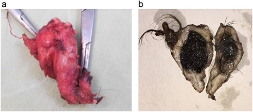
Under general anesthesia, a modified Blair incision was given, and dissection was done to identify the cyst. Intraoperatively, the cyst was found to be attached to the cartilaginous external auditory canal by a stalk with a tuft of hair. Stalk was excised from the attachment, and the cyst was removed along with the superficial lobe of the parotid gland. The facial nerve was identified and preserved. Postoperatively, the patient recovered well with no signs of facial nerve injury. On pathological examination, the excised specimen measured 4.5 × 3 × 2 cm. The cyst had a thick gray-white wall. The lumen was filled with hair and pultaceous material (Fig. 2). On microscopic examination, the cyst wall was lined by keratinized stratified squamous epithelium, and most of the cyst wall showed granulation tissue, foreign body giant cell reaction, numerous hair shafts and adjacent normal salivary gland lobules separated from the cyst by a thick fibrous wall. The features described were consistent with the diagnosis of dermoid cyst. The postoperative period was uneventful, and the patient was currently under follow-up for 1 year.

(a) Excised specimen (b) cut section showing the cyst filled with hair and pultaceous material.
DISCUSSION
Dermoid cysts are rare, lined by epithelium and enclosed in a sac that contains tissues of both mesodermal and ectodermal origin. The most common regions of dermoid cysts are head and neck regions involving nasal, orbital and oral (submental and submaxillary) regions. Since many embryonic structures fuse in these areas, their regions are more prone to develop dermoid cysts [3, 4]. The parotid glands are a common site to get affected by cysts and congenital lesions [6]. A cystic lesion can occur in any portion of the parotid gland. Dermoid cysts in adults are uncommon, and involving the parotid region is extremely rare, with only a few cases reported.
Dermoid cysts are further classified into the following three categories on the basis of pathogenesis and microscopic appearance [5].
(i) Congenital dermoid cysts of the teratoma category.
(ii) Acquired dermoid cysts.
(iii) Congenital inclusion dermoid cysts.
Choi et al. described that it is difficult to classify the dermoid cyst of the parotid gland using the above classification. They hypothesized that they could be classified under the third category (congenital inclusion dermoid cysts), whereas Yigit et al. attributed them to the second category (acquired dermoid cysts) [6, 7]. Differential diagnosis of parotid dermoid cyst includes tumors like epidermoid cysts, lipoma, neurofibroma, teratoma, pleomorphic adenoma of the salivary gland, Warthin’s tumor and mucoepidermoid tumor [4, 6].
Clinically, it is difficult to diagnose the parotid dermoid cyst definitively since, on physical examination, it has no characteristic findings [6]. In our case, all the preoperative investigations suggested a cystic lesion. Since there was a history of a dermoid cyst excision, we suspected it to be a recurrent parotid dermoid cyst. Dermoid cysts are generally encapsulated, which facilitates dissection. Dwivedi et al. [8] reported a case of dermoid cyst for which enucleation was sufficient. Superficial parotidectomy is common surgical management for the parotid dermoid cyst. Total parotidectomy is performed in rare cases of dermoid cysts, i.e. cysts located in deeper parts of the parotid gland [7]. The risk of facial nerve involvement with a parotid dermoid cyst should be considered during surgical planning, and nerves should be dissected carefully from the cyst to prevent damage to the nerve and further neurological deficits. Choi et al. [6] described two cases of parotid masses, which were previously operated but had recurred. The first described case underwent simple excision previously, whereas the second had a history of an aspiration biopsy, but both cases had a recurrence of the mass; hence superficial parotidectomy was performed. Even in our case, simple cyst excision was done previously, but it did not suffice, leading to the recurrence of the dermoid cyst; hence superficial parotidectomy was done, taking utmost care to preserve the facial nerve. Postoperatively, there was no recurrence and no facial palsy in our case, even at a follow-up of 1 year.
CONCLUSION
Although intraparotid dermoid cysts are rare, they must be considered as a differential diagnosis in tumors of the parotid region, which are painless, recurrent and have a soft consistency. Proper history taking, examination and preoperative diagnostic workup must be made to plan for appropriate management. The final diagnosis should always be made by histopathological examination. In recurrent cases, superficial parotidectomy is the treatment of choice.
ACKNOWLEDGEMENTS
The authors would like to thank the patient and attendants for accepting for publication. Sincere thanks to Squad Medicine and Research (SMR) for their support and guidance.
CONFLICT OF INTEREST STATEMENT
None declared.
FUNDING
The author(s) received no financial support for research, authorship and/or publication of this article.
RESEARCH INVOLVING HUMAN PARTICIPANTS AND/OR ANIMALS
This case report involves a human participant. All procedures performed in the study involving human participant were in accordance with the ethical standards of the departmental research committee.
INFORMED CONSENT
Written informed consent was obtained from the participant regarding submission of case report, publishing their data and photographs to the journal.
AUTHORS' CONTRIBUTIONS
George KS – conceptualization, formal analysis, investigation, writing (review and editing), project administration. Sandeep R – conceptualization, investigation, writing (review and editing), supervision. Ramji KVV – writing (review and editing), project administration. Kuchana KS – investigation, writing (original draft, review and editing). Suvvari TK – writing (original draft, review and editing). Thomas V – writing (original draft, review and editing), supervision.
References


