-
PDF
- Split View
-
Views
-
Cite
Cite
Qusai Mashlah, Ali M Zaher, Hajar Odah Bashi, Hanady Zwaraa, Husam Dalati, Torsion of the whole ileum and hemorrhagic necrosis because of a giant Meckel’s diverticulum in a 9-month-old child, Journal of Surgical Case Reports, Volume 2023, Issue 6, June 2023, rjad336, https://doi.org/10.1093/jscr/rjad336
Close - Share Icon Share
Abstract
Intestinal ischemia caused by torsion of a freely hanging Meckel’s diverticulum (MD) resulting in the need for resection is an uncommon complication. We present an extraordinary case of a 9-month-old male with acute abdominal symptoms because of intestinal ischemia and necrosis that necessitated resection of the entire ileum. This was caused by torsion around a particularly large MD.
INTRODUCTION
Meckel’s diverticulum (MD) is the most prevalent congenital anomaly of the small intestine, with a lifetime risk of 4–6% for developing complications. Clinical symptoms and complications may arise from small bowel obstruction, bleeding, inflammation, umbilical abnormalities or neoplasia. Intestinal obstruction is the most common surgical finding in symptomatic patients, accounting for 41% (with a range of 14–86%) of cases resulting from MD [1]. MD can lead to bowel obstruction via various mechanisms, such as intussusception, volvulus, vitelline bands/remnants, incarcerated Littre hernia and others [2]. Intussusception is slightly more common in children, and volvulus in adults.
CASE REPORT
A 9-month-old male with sickle cell disease presented with severe vomiting, feeding intolerance, irritability and retention of stool and gasses for over 24 h. The patient did not have a history of rectal bleeding. On clinical examination, the patient exhibited mild abdominal tenderness. An ultrasound scan (US) showed a small to moderate amount of free fluid in Morrison’s sinus and splenic sinus, as well as between intestinal loops. No visible signs of intussusception were observed. The patient’s laboratory results showed a white blood cell count of 5400, hemoglobin level of 10.5, platelet count of 120 000, CRP of 6, Na of 138 and K of 5.4.
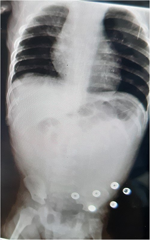
An abdominal X-ray revealed no gas liquid levels or free gas in the abdomen. Intestinal loops were dilated, and there was a lack of gas in the abdomen and pelvis. Twelve hours later, the patient developed severe abdominal pain with generalized abdominal guarding. The hemoglobin level dropped to 6.8, necessitating an emergency exploratory laparotomy (Fig. 1).

AXR: no gas liquid levels—no free gas in the abdomen—dilation of intestinal loops and lack of gas in the abdomen and pelvis.
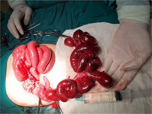
During surgical investigation, a small bowel torsion was discovered extending from the ileocecal junction to 120 cm of ileum, which included a broad-based, inflamed giant MD. Attempts to reverse the torsion and warm the intestines for 25 min did not improve perfusion. As a result, the damaged intestine, cecum and appendix were amputated (Fig. 2), and an end-to-end two-layer ileocolic anastomosis was performed.

Image during resection showing giant MD and necrotic small intestine.
The patient began oral feeding on the 5th day post-surgery with good tolerance and was discharged on the 7th day. Histopathological examination revealed hemorrhagic necrosis in the intestine up to the ileum margins, consistent with torsion of the intestine with diverticular formation showing hemorrhagic necrosis. The appendix showed nonspecific follicular hyperplasia with bleeding.
DISCUSSION
Approximately 4% of individuals with MD will exhibit symptoms. The predominant causes of symptomatic Meckel’s are intestinal obstruction, which can occur through various mechanisms such as intussusception or volvulus. Volvulus can arise from fibrous or vascular remnants of the omphalodiverticular tract (extending from the tip of the diverticulum to the abdominal wall). Giant Meckel diverticula or vitelline cysts may also cause volvulus. If the volvulus causes ischemia, the patient may exhibit signs of peritonitis and may present in severe cases such as the one being discussed. Torsion of an MD resulting in intestinal ischemia that requires resection is a rare complication [3]. In rare instances, necrosis of the intestine because of MD with no identifiable point of fixation has been reported [1, 4–7].
Acute midgut volvulus (MGV) is considered the most severe form of volvulus and has been associated with a fourfold increase in mortality risk in comparison to segmental volvulus. The overall mortality rate for MGV ranges from 2.2 to 16%. Approximately 75% of MGV cases occur in the 1st year of life, and most cases occur during the neonatal period. The incidence of bowel necrosis with MGV in other reports was found to range from 2.9 to 48%. Heart rate; mean arterial pressure; pH; and serum CRP, sodium and albumin levels are independent predictors for intestinal ischemia and necrosis in patients with MGV [8]. The presence of superior mesenteric artery collapse, ascites and a large intestinal twist on US imaging were significant predictors of intestinal ischemic changes [9].
When the ileum adjacent to the diverticulum is ischemic, resection of the affected bowel with an ileo-ileostomy is preferred [5]. In patients with intestinal obstruction, delaying surgery for more than 36 h increases the mortality rate from 8 to 25% [10].
CONCLUSION
The occurrence of intestinal ischemia necessitating resection because of MD is an infrequent yet life-threatening complication. Prompt diagnosis and timely surgical intervention are crucial in preventing necrosis of the midgut, mortality and short bowel syndrome resulting from extensive small bowel resection. This case reinforces the possibility of bowel torsion in MD without the presence of a fibrous band or adhesion. Furthermore, laboratory analyses and abdominal imaging with ultrasound may not reveal any evidence of ischemia or necrosis in the small bowel.
CONFLICT OF INTEREST STATEMENT
None declared.
FUNDING
None.
DATA AVAILABILITY
All data underlying the results are available as part of the article and no additional source data are required.


