


Abstract
A 22-year-old female presented to our clinic with the redevelopment of compressive symptoms after undergoing right carpal tunnel release 2 years prior. During revision surgery, the application of a chitosan polysaccharide membrane wrap over the median nerve was secured with three hemoclips. Follow-up revealed that these clips traveled distally to the volar aspect of the patient’s small finger necessitating excision. Hemoclips in hand surgery serve as a useful tool for securing constructs; however, the recommendation to use them sparingly is made because of the potential for migration and exacerbation of symptoms.
Introduction
After their initial introduction, endoscopic clips (‘hemoclips’) developed as a primary method for ligating vessels and achieving hemostasis in abdominal or variceal surgeries [1–3]. Because of their broad applicability and ease of use, these clips have found niche functions in fields such as cardiac surgery, neurosurgery, urology, and plastic surgery [4–8]. Rare complications, such as clip migration, have been sparsely noted within the literature of laparoscopic procedures since 1992 [9]. Clip migration in the general surgery literature has largely been relegated to case reports [9]. Noted symptoms caused by migration have included obstructive jaundice, gastrointestinal bleeding with anemia, fistula formation, choledocholithiasis, or duodenal ulcer development [10–12]. These symptoms have ranged in presentation from 2 weeks up to 35 years postoperatively [11]. Steps to transition from metal clips to nonabsorbable polymer locking clips (Hem-o-lok clips) have been made in hopes of combatting migration, but have been largely unsuccessful [2, 10, 13, 14].
Although a large amount of data from the general surgery literature has been gathered, none has been documented regarding their usage within the realm of Orthopedic Surgery. This report presents a case where a patient developed an exacerbation of symptoms after undergoing a revision carpal tunnel release (CTR) with application of hemoclips to secure a membrane nerve wrap. The patient provided their written informed consent in the publication of this report.
Case report
A 22-year-old female with a relevant medical history of Type 2 diabetes and hypertension initially presented to our clinic with complaints of right-hand pain radiating from her fingers to her elbow. The patient had undergone a prior CTR on the ipsilateral side 2 years prior, with no interventions on the contralateral side despite similar complaints of compressive symptoms. On exam, the patient demonstrated a positive Phalen’s test and Tinel’s sign with 4/5 strength on resisted opponens testing.
Repeat electromyography (EMG) and monofilament testing demonstrated some continued compression at the carpal tunnel and attempted nonoperative management included night splinting, physical therapy, and a corticosteroid injection (Fig. 1). The patient continued to vocalize complaints of right thumb numbness, sharp pain in her index finger, and an inability to distinguish temperature; therefore, she was subsequently scheduled for a revision CTR.
![Semmes Weinstein testing values after redevelopment of right-hand symptoms [17].](https://oupdevcdn.silverchair-staging.com/oup/backfile/Content_public/Journal/jscr/2023/10/10.1093_jscr_rjad548/1/m_rjad548f1.jpeg?Expires=1785870855&Signature=hEcPBtElgSX-7-x638KbqMdLbsdHjTJECEnZKtrdULokt~W~Q1QU1PPu674QXISr6Jj24iVVpfLQStEFwbM0j0kJBlMgueiCkIYuMuuoRvg~L-hY-ghHS0CUp6DsgYoWdQGr-P79xJzpUK6ztkrELG7jbvQj5MukU9vG9PnUAryfpcJni7EujToun1uxMfw-rR5x6KDIIoCt3Vxeb~aVoW-328BERp2eJ2rCOiELWKG9Qu-fMPFEmlEqExwI4m3trPYtpnb~LfIsmCNJAtvFPZLd9z5LUdK7pQCwczH00IQ7MAxpMT2x~xbqDz6N3FaZjAKOytcX~njktsyvRuw3~g__&Key-Pair-Id=APKAIYYTVHKX7JZB5EAA)
Semmes Weinstein testing values after redevelopment of right-hand symptoms [17].
In the operative theater, her previous skin incision was utilized to prevent scar contracture. A thickened amount of scar tissue at the proximal portion of the carpal tunnel was identified. Her median nerve had scarred to the underside of the retracted radial leaflet of the transverse carpal tunnel ligament. Copious scar tissue was removed to free the nerve and an internal neurolysis was completed. The nerve was traced distally to the recurrent motor branch in the thenar musculature to ensure no damage to the neural structures. Neuroshield® (a chitosan polysaccharide membrane wrap) was wrapped around the nerve and secured with three hemoclips followed by Tisseel glue (a fibrin sealant) to protect the median nerve (Fig. 2).
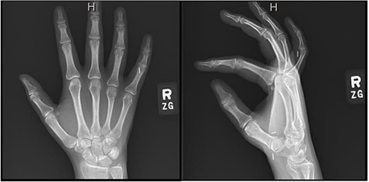
Right hand imaging 1-month status post revision CTR demonstrating hemoclip placement with initial asymptomatic migration of a single clip to the small finger proximal interphalangeal joint.
Four months postoperatively, the patient presented with complaints of pain at her small finger distal interphalangeal joint, and verbalized feeling small staples at the symptomatic site. Radiographic imaging demonstrated that the three previously placed hemoclips had migrated along the flexor tendon tract to the volar aspect of her right small finger proximal interphalangeal joint (Fig. 3). Surgical excision of the clips involved utilizing a Bruner-style incision volarly followed by a small window into the tendon sheath. One hemoclip was identified at the level of the proximal interphalangeal joint sandwiched between the flexor digitorum profundus and the superficialis tendons. Two more clips were identified proximally.
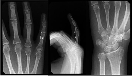
Right hand imaging 4-months status post revision CTR demonstrating symptomatic triple hemoclip migration into the volar aspect of the small digit.
Two weeks postoperatively from clip excision, the patient’s thumbs up, OK, and two to three cross exams were intact. Sensation was intact at the medial, ulnar, and radial distributions. At a 2-month follow-up from her excision or 1-year follow-up from the original revision, the patient’s incisions had healed well without concerns for infection or drainage, and she remained neurovascularly intact. Her small finger and original compressive symptoms had resolved without complications.
Discussion
Hemoclip migration was first noted in 1992 by Raoul et al. after laparoscopic cholecystectomy procedures, with this number growing to over 100 cases in the past decade [2, 9, 13]. In gastrointestinal surgery, migration down the biliary tract or into the duodenum has been documented to lead to an array of symptoms including obstructive jaundice, gastrointestinal bleeding with anemia, fistula formation, choledocholithiasis, or duodenal ulcer development [10–12]. The general surgery literature has described specific technical reasons for clip failure, but many generalizable nonspecific causes have been documented as well. Multiple authors have observed that a localized postsurgical inflammatory process may lead to erosion of the surrounding structures and eventual clip migration [10, 12–14]. In a similar fashion, a larger number of clips applied are often directly proportional to the likelihood of migration occurring [3, 7, 14]. Most notable, however, has been the discussion that the placement of a clip induces a latent inflammatory-rejection response by the host [2, 3, 13–15]. While plausible as to why there have been delayed presentations of up to 35 years, our case presented within 4 months. The symptoms presented by our patient can be assumed to be largely because of the presence of the clips in the tendon sheath at the level of the proximal interphalangeal joint instead of an underlying chronic inflammatory process.
As mentioned previously, there have been movements to transition from metallic clips to more inert polymers. Mozafari et al. [16] found that allergies secondary to hemoclip placement may cause type IV hypersensitivity symptoms most commonly because of the composition of nickel or titanium in metallic clips. The patient in our case received metal clips and if unnoticed, this may have developed into a larger acute inflammatory process. There is no standardized regulation of allergy testing before procedures and given the high prevalence of metal allergies within the general population at 10%–15%, it remains questionable whether the convenience of using hemoclips versus other materials is beneficial [16].
Although the present case differs from hemoclips being used for their traditional hemostatic usage, the migration that occurred was concerning as the patient developed symptoms shortly after their placement. Clip migration could be a nidus for structural damage within the hand or lead to an allergic or inflammatory response. Minimalistic usage of these clips is recommended as there is potential for complications and further surgical intervention. Further data and recommendations are needed to assess the implications of uncommon cases where migration can occur and lead to symptom development.
Conflict of interest statement
The authors have no conflicts of interest to disclose.
Funding
This research received no funds, grants, or other support from agencies in the public, commercial, or not-for-profit sectors.
Data availability
Access to the data supporting the case report is restricted due to the Health Insurance Portability and Accountability Act. Deidentified data can be provided by the authors upon request.
