-
PDF
- Split View
-
Views
-
Cite
Cite
Sarah McLain, Jack Cecire, Allan Mekisic, Incarcerated inguinal hernia containing urinary bladder and bladder calculi, Journal of Surgical Case Reports, Volume 2023, Issue 1, January 2023, rjad010, https://doi.org/10.1093/jscr/rjad010
Close - Share Icon Share
Abstract
Inguinoscrotal herniation of the bladder is an uncommon condition. Clinicians should have a high index of suspicion for involvement of the bladder within a hernia when the lower urinary tract urinary symptoms are concurrently present in patients presenting with a painful groin lump. We report the case of a patient who presented with an inguinoscrotal hernia involving the urinary bladder, which was irreducible due to the fact that the herniated portion of the bladder contains multiple containing calculi. The patient underwent successful operative repair of the hernia and had an uncomplicated post-operative course. Key takeaway messages for clinicians are that the presence of lower urinary tract symptoms in a patient concurrently presenting with a groin lump should raise suspicion of bladder involvement within the hernia and that pre-operative imaging is valuable to identify the contents of the hernia to allow safe operative planning and to reduce the risk of bladder injury during surgery.
INTRODUCTION
Inguinoscrotal herniation of the bladder is an uncommon condition. The patient may present with a painful groin lump and can be simultaneously experiencing lower urinary tract symptoms or urinary retention [1]. Clinicians should have a high index of suspicion for involvement of the bladder within an inguinoscrotal hernia in such presentations. Pre-operative imaging with computed tomography (CT) scanning allows for the identification of the bladder involvement, facilitates operative planning and can reduce the risk of bladder injury during surgery [2, 3]. We report the case of a patient who presented with an incarcerated inguinoscrotal hernia involving the urinary bladder and containing bladder calculi.
CASE PRESENTATION
A 90-year-old male presented to the emergency department of an Australian metropolitan tertiary hospital with a 24-hour history of a painful right inguinal swelling and complaints of poor drainage of urine via his long-term indwelling urinary catheter and leakage of urine around the catheter. He had a known right-sided inguinal hernia that had been present for the past 20 years, which had always been reducible and caused minimal discomfort. He was not experiencing any symptoms of a bowel obstruction, with no abdominal pain, nausea or vomiting. His bowels were opening, and he was passing flatus.
The patient had not had any previous abdominal surgery. His urological history was significant for locally advanced prostate cancer diagnosed 11 years earlier and was managed non-operatively with androgen deprivation therapy (leuprorelin acetate depot injection of 22.5 mg every 3 months) and a long-term indwelling urinary catheter changed at six weekly intervals due to urinary retention secondary to prostatic enlargement. Four years prior, he had undergone cystoscopy and laser lithotripsy to remove a 11 × 8 mm bladder calculi.
On examination in the emergency department, his vital signs were normal and he was afebrile. His body mass index was 27. His abdomen was soft, not distended and non-tender. There was 3 × 3 cm firm right inguinal lump, which was tender on palpation and not reducible, with no overlying skin changes. Blood demonstrated normal renal function, electrolytes and inflammatory markers. His urinary catheter was exchanged for a new 16 Fr Foley catheter without difficulty.
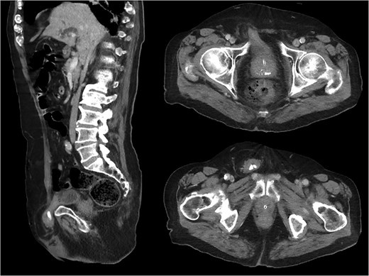
CT abdomen and pelvis was performed to assess whether any bowel was involved in order to determine the urgency of surgical intervention. The CT showed a right-sided inguinal hernia containing a portion of the urinary bladder, with bladder calculi entrapped within the hernia (Fig. 1).

Sagittal and axial CT images demonstrating right-sided inguinal bladder hernia containing radio-opaque bladder calculi.
The patient was admitted under the care of the general surgeon and was booked for theater after providing informed consent for open repair of inguinal hernia. Surgery took place the following morning as there were no signs of strangulation (clinically or on imaging). A right groin incision was made and the external oblique opened to the external ring. The spermatic cord was mobilized, commencing at the pubic tubercle. The ilioinguinal nerve and cord structures were identified and preserved. A direct hernia was noted, with palpable bladder calculi preventing its reduction. The hernia sac was not opened due to the risk of iatrogenic bladder injury. The defect in the posterior wall of the inguinal canal at the neck of the hernia had to be extended in order to facilitate reduction. The transversalis fascia was plicated to maintain reduction. A Lichtenstein repair was performed, and the posterior wall of the inguinal canal was reinforced with a partially absorbable lightweight mesh (poliglecaprone-25/polypropylene). The Lichtenstein mesh repair is routine in our institution, with evidence supporting a lower risk of hernia recurrence [4]. The mesh was secured inferiorly from the pubic tubercle along the inguinal ligament in a continuous fashion and superiorly to the transversalis fascia with interrupted polypropylene sutures. The external oblique was closed, followed by closure of the wound in layers.
The patient had an uncomplicated post-operative course. The following morning, he was clinically well, mobilizing comfortably and his urinary catheter was draining well. He was discharged home with instructions to avoid strenuous activity and heavy lifting for the next 6 weeks. At follow-up 8 weeks post-operatively he remained well, had no recurrence of the hernia and surgical wound had healed.
DISCUSSION
Inguinal hernia containing bladder is an uncommon presentation, frequently quoted at rates of 1–5% of all hernias [1–3]. Risk factors include older age, male sex, obesity, weakened abdominopelvic musculature and chronic bladder outlet obstruction, with resulting enlargement of the urinary bladder causing it to come into proximity of the hernia orifices [1, 5]. Although inguinal bladder hernia is often described as an asymptomatic incidental finding, general surgeons should have a high index of suspicion when patients present with an inguinal hernia and associated lower urinary tract symptoms. Cases described in the literature include symptoms such as difficulty initiating urination, double voiding and incomplete micturition [1, 3, 5, 6]. Some cases presented with urinary retention, obstructive uropathy with acute renal failure and sepsis from urinary tract infection [2, 3, 5]. To our knowledge, this is one of the first reported cases in the literature where inguinal bladder hernia was present in a person with long term urinary catheter and with bladder calculi preventing reduction of the hernia.
The literature often reports inguinal bladder hernias are commonly diagnosed intra-operatively rather than pre-operatively, increasing the risk of damage to the bladder when it is an unexpected finding at the time of surgery [6]. Pre-operative imaging with CT is therefore helpful to allow for procedural planning and prevent bladder injury [2, 3].
Inguinal bladder hernia is an uncommon presentation, and the key learning points from this case are that it should be suspected in patients presenting with an inguinal hernia and urinary symptoms. Pre-operative imaging is highly beneficial to identify the contents of the hernia and to avoid iatrogenic bladder injury at surgery. In this patient, the urinary calculi in the herniated portion of the bladder led to the hernia becoming irreducible and required the surgeon to address this at operation by extending the defect in the posterior wall of the inguinal canal in order to facilitate reduction.
CONFLICT OF INTEREST STATEMENT
None declared.
FUNDING
None.
DATA AVAILABILITY
There is no additional data outside of what is included in the manuscript.


