


Abstract
Tessier no. 7 clefts are characterized by macrostomia, facial muscular diastasis and maxillary and zygomatic bone abnormalities. It is caused by a lack of ectomesenchyme formation or penetration of the maxillary and mandibular processes during the fourth and fifth weeks of development. A case of bilateral transverse facial cleft with an accessory maxilla and an osseous choristoma is presented. The diagnosis of accessory maxilla was based on clinical findings due to the inaccessibility of orthopantomography and computed tomography scan. Orbicularis oris muscle reconstruction, cheiloplasty and excision of accessory maxilla were done. Histopathological examination of the bony lesion showed an osseous choristoma. There were no postoperative complications or local recurrence of the lesion excised. This case report demonstrates the importance of early diagnosis and intervention in maxillofacial congenital anomalies. Cheiloplasty restores function and gives the patient a natural appearance. The excision of accessory bone prevents further complications in the child’s growth.
INTRODUCTION
Craniofacial clefts are rare congenital anomalies. Their cause is unknown, but they are associated with genetic predispositions and environmental factors [1]. The Tessier clefting system is a common classification method based on descriptive and anatomical characteristics, giving numbers from 1 to 30 to different clefting sites based on how their anatomy connects to the sagittal midline [2].
Tessier no. 7 or transverse facial clefts are characterized by macrostomia, facial muscular diastasis and maxillary and zygomatic bone abnormalities [3]. It may occur alone or as part of a syndrome. It is caused by a lack of ectomesenchyme formation or penetration of the maxillary and mandibular processes during the fourth and fifth weeks of development, resulting in the failure of the maxillary and mandibular processes to fuse at the first pharyngeal arch and a cleft at the commissures of the lips. The deep muscles appear to be split [4, 5]. A sulcus at the commissure may be the only finding [6, 7]. It usually goes 1–2 cm past the front edge of the masseter, but in more severe cases, it can go all the way to the ear and beyond [3, 8, 9].
Tessier no. 7 clefts may be associated with additional anomalies, such as a maxillary or mandibular accessory, a rare clinical entity characterized by the presence of extra bone lying posterior to the tuberosity of the maxilla caused by abnormal growth of the zygomatic arch [10]. Patients also frequently exhibit preauricular tags and sinuses. Transverse facial clefts are more common in males than females. The incidence is between 1 in 60 000 and 1 in 300 000 live births [6, 11, 12].
A case of bilateral commissural clefts with the presence of skin tags and a left accessory maxilla in a woman is discussed.
CASE REPORT
A 24-year-old woman brought her 1-month-old daughter to the Dental Unit at Ho Teaching Hospital because she found a hard mass in her daughter’s mouth during breastfeeding and she was also concerned about the shape of the baby’s mouth. The mass has slightly increased in size as the child ages, and it produces irritation in her breast. There was no family history of maxillofacial malformations. The baby was born through a eutocic delivery.
Extraoral examination revealed a depression on the right side of the face that extended from the commissure to the external cantus of the eye, bilateral commissural clefts and skin tags. There was a slight deviation of the mouth to the left. The midline of the lower lip shifted to the left. The left commissural cleft was pulled laterally and downward. The left zygomatic region was more prominent than the right (Fig. 1).
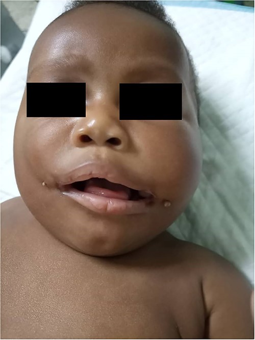
Preoperative picture of the patient with bilateral commissural cleft.
Intraorally, a round shaped mass was found in the left cheek adjacent to the commissural cleft, about 1 cm in diameter covered by a thin and pale buccal mucosa; bony hard on palpation with an equally hard submucosal continuity which ends in the posterior aspect of the maxilla producing stretching of the cheek and the lower lip. Mouth opening was adequate. A diagnosis of bilateral commissural cleft and left accessory maxilla was done. The full blood count, abdominal ultrasound and echocardiography were normal. A panoramic X-ray and computed tomography (CT) scan were requested but not available.
Orbicularis oris muscle reconstruction, cheiloplasty using a mucocutaneous flap, Z-plasty and excision of commissural tags and accessory maxilla were done under orotracheal general anesthesia. Surgery was started from the right site; various points for the design of the commissural mucocutaneous flaps were marked (Fig. 2). Mucocutaneous flap was raised, keeping its base in the buccal mucosa (Fig. 3). Reconstruction of the new commissure was done suturing the mucocutaneous joint of the upper and lower lips. Subsequently, small incisions were made in the distal part of the mucocutaneous flap to remove the skin tag. After the closure of the intraoral cleft, the orbicularis oris muscle was identified, exposed and sutured. A Z-plasty was performed; the two triangular flaps were created, transposed and sutured. The cleft on the right site was closed, after that we proceeded to the left, beginning with the removal of the bony lesion in the cheek where a linear incision was made and the bone was dissected and detached posteriorly to the maxilla (Fig. 4). The closure of the left commissural cleft was done following the same principles applied to the right site (Fig. 5). Daily dressing was done, and sutures were removed after 10 days.
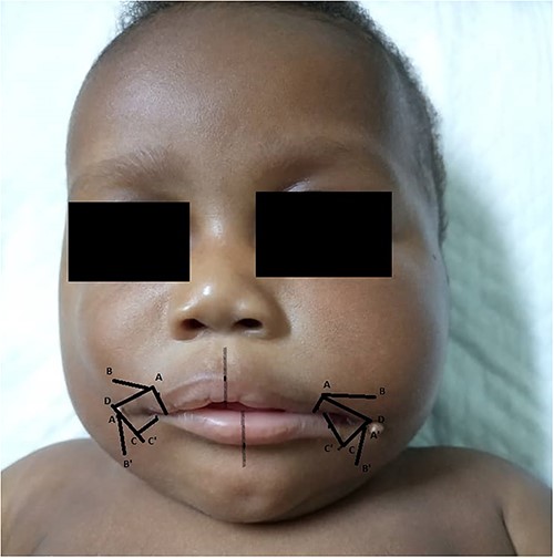
Graphic design of the commissural mucocutaneous flaps.
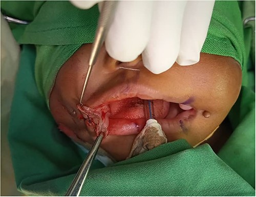
Mucocutaneous flap raised keeping its base in the buccal mucosa.
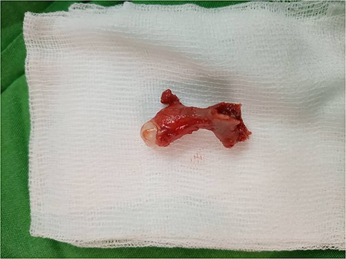
Accessory maxilla removed from the left site with a presence of a distal lesion.
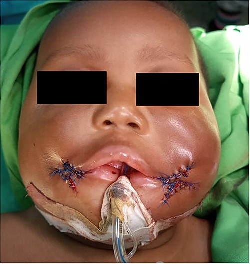
Z-plasty of both commissural cleft.
The bony lesion was taken for a biopsy: the histopathological report described sections showing relatively normal squamous epithelium overlying a thick coat of fibroconnective tissue containing islands of epithelial structures. There were multiple bony trabeculae in loose fibrovascular connective tissue. Elsewhere, bone with normal marrow was noted. Diagnosis of osseous choristoma was made.
Regular follow-ups have been done for a year, the wounds have healed satisfactorily, and there were no postoperative complications or local recurrence of the lesion excised (Fig. 6).
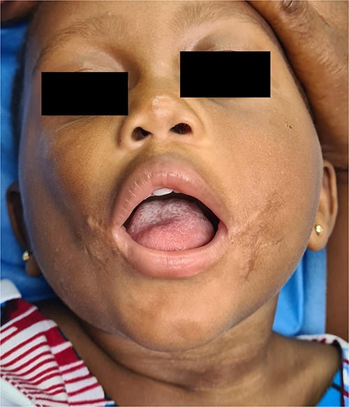
Facial appearance of the patient’s face 1 year after surgery.
DISCUSSION
A case of a child with bilateral commissural clefts and an accessory maxilla was presented, which was neither diagnosed during prenatal examinations nor at birth, rather it was the discomfort in the mother’s breast during the feeding the main reason for bringing the baby for consultation, despite the evident deformity on the baby’s lips and face. It is important to examine fetal craniofacial structures during prenatal examinations and the oral cavity of the infant after birth for an early diagnosis of malformations and treat them at the appropriate time [13]. Orthopantomography and CT scan are very useful in the diagnosis of the bony abnormalities associated with Tessier no. 7 cleft; in low-income countries like Ghana, these complementary tests are not available in all the regions; in our case, they were not accessible and the diagnosis was made based on clinical features.
Cases of bilateral commissural clefts with an accessory maxilla are uncommon. In a literature review conducted by Sun et al., 24 patients with Tessier no. 7 cleft were reported from 2000 to 2020; the most common clinical manifestation of Tessier no. 7 cleft was bilateral facial clefts. Only three patients exhibited unilateral accessory maxilla [14]. In the literature reviewed, there was not any report of osseous choristoma associated to these kinds of anomalies. The most common site for oral osseous choristoma is the posterior third of the tongue. Localization of these lesions in oral cavity sites other than tongue is relatively uncommon [15].
The objectives of the commissural cleft repair are to achieve a symmetrical mouth opening, to make the commissures appear natural and to maintain the function of the orbicularis oris muscle [16]. Many surgical techniques have been described for the closure of a commissural cleft in order to achieve these objectives [6]. In this case, a Z-plasty was performed because it is relatively easy to design and reduce wound contraction.
CONCLUSIONS
This case report demonstrates the importance of early diagnosis and intervention in maxillofacial congenital anomalies. A thorough intraoral examination helps with the diagnosis even when imaging tests are not available. Repositioning the orbicularis oris muscle and Z-plasty during the closure of the bilateral commissural clefts restores the function of the lips and the contours of the mouth giving the patient a natural appearance with minimal wound contraction. The excision and biopsy of the bony lesion prevent further complications in the child’s growth.
CONFLICT OF INTEREST STATEMENT
None declared.
FUNDING
The authors did not receive financial support for the research and publication of this case report.
