-
PDF
- Split View
-
Views
-
Cite
Cite
Botaitis Sotirios, Deftereos Savas, Zachou Maria-Eleni, Vasileiadis Stauros, Perente Sempachedin, Isolated echinococcal infection of the spleen: a rare case report, Journal of Surgical Case Reports, Volume 2022, Issue 6, June 2022, rjac217, https://doi.org/10.1093/jscr/rjac217
Close - Share Icon Share
Abstract
Echinococciasis is a zoonotic infection observed in almost every anatomic location in the body. The liver and lungs are the most frequent sites of infection. However, spleen involvement is rare, and isolated splenic hydatid disease is even less common. Herein, we report a case of isolated splenic hydatid cyst without any tissue involvement that was treated with the classical surgical approach.
INTRODUCTION
Echinococciasis, also known as hydatid disease (HD), is a zoonotic infection caused by the larvae of Echinococcus in the genus Echinococcus. The liver (70%) and lungs (15–47%) are the most common sites of the disease. Meanwhile, it is less frequently noted (15%) in the other body parts [1, 2]. Primary splenic hydatidosis is characterized by spleen involvement in the absence of diseases in other organs and is an uncommon manifestation of HD. Splenic hydatid cysts may occur as a part of disseminated disease or may be isolated. Herein, we report a case of isolated splenic hydatid cyst.
CASE REPORT
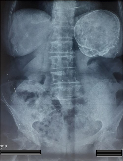
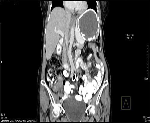
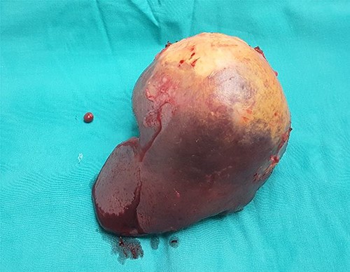
A 74-year-old woman was admitted to the internal medicine ward due to cough, pain and heaviness in the left upper abdominal quadrant. Pain was localized to the left upper abdominal quadrant, and it intensified within the last 2 months. The laboratory test results were normal. However, physical examination revealed tenderness upon palpation in the left upper quadrant and splenomegaly. Moreover, thoracic and abdominal radiography revealed an egg shell-like calcified well-circumscribed ovoid mass at the left upper abdominal quadrant (Fig. 1). Computed tomography (CT) scan of the abdomen revealed a 10.5 × 9.6 × 9.5-cm ovoid splenic cyst with peripheral rim calcification. The cyst had homogeneous internal substance with no prominent daughter cysts or scolices (Fig. 2). Thoracic and abdominal CT-scan did not reveal other pathologies and cystic lesions. The patient underwent exploratory laparotomy. Then, a splenic cyst was observed, and the spleen was found to be densely adherent to the diaphragm and gastrosplenic ligament. A laparotomy was performed and the spleen with the cyst was removed without opening. A well-described accessory spleen with a diameter of 8 mm was found in the gastrosplenic ligament. The splenectomy specimen and the accessory spleen weighed 320 g and 4 g, respectively (Fig. 3), and they were sent for histopathological examination. The patient’s postoperative recovery was uneventful. She was discharged on the sixth postoperative day and treated with albendazole for 3 months.
DISCUSSION
Splenic HD condition, which accounts for 2.5–5.6% of all abdominal HDs, can appear at all age groups and in both sexes, and they are commonly solitary [3]. In up to 30% of cases, it may be associated with cysts in other body parts [4, 5]. The parasite may reach the spleen via the blood stream, lymphatic system [6] and via reflux into the spleen from the portal vein during increased intra-abdominal pressure [7]. Secondary splenic HD can originate from either rupture of hydatid cyst in the intraperitoneal organs or by systemic dissemination [8].

Abdominal X-rays with a calcified cyst at the left upper quadrant of the abdomen.

CT of the abdomen shown a 10.5 × 9.6 × 9.5-cm ovoid splenic cyst with peripheral rim calcification.

The symptoms of splenic hydatid cysts are nonspecific, and based on the size, location and compulsive effects of the cyst which primarily cause abdominal burden in the left hypochondrium or epigastrium. Approximately 30% of splenic cysts are silent. A plethora of symptoms have been described, and these include nausea, weight loss, dull pain, bloating, dyspepsia, constipation, lumbar discomfort, intermittent fever, jaundice, cough and dyspnea [1]. Due to the slow rhythm of progression (0.3–1 cm per year) and the absence of symptoms or direct parasitological evidence, echinococcosis cyst is frequently discovered incidentally while investigating other diseases [9].
The main problem in the diagnosis of splenic hydatidosis is differentiating it from other splenic lesions. A more definitive diagnosis can be obtained via abdominal radiography and ultrasonography and CT scan. A simple radiography of the abdomen can identify egg shell-like calcification in the spleen that strongly indicates the presence of splenic hydatidosis, as our patient. On abdominal ultrasonography, splenic hydatid cysts may present as a single or, rarely, multiple anechoic spherical cystic lesions with well-defined limits that can be hyperechoic due to calcifications. Abdominal CT scan is the most sensitive procedure for diagnosing such lesions. Moreover, it can validate the presence of hydatid cyst and can be performed to assess the anatomical relations of the cyst with adjacent viscera before surgery.
The complications of splenic hydatid cysts arise from intra-abdominal organ compression, adhesions with the adjacent organs, fistula formation into hollow viscera such as the colon and stomach, intra-abdominal rupture of the cyst, secondary infection of the cyst, hypersplenism, sympathetic pleural effusion, severe urticaria, calcification, splenic atrophy [1]. In our patient, ring calcification with spleen atrophy and an accessory spleen were observed.
Surgery is the common treatment of choice, and the surgical approach is based on the number, location and size of cysts. In patients with multiple cysts in different initial organs and particularly in the peritoneal cavity, surgery is contraindicated, and treatment with antihelminthics as albendazole is preferred [10]. The surgical procedures for HD include total or partial splenectomy and spleen-preserving surgery, using the conventional or laparoscopic approach. Single peripheral and small inactive or superficial cysts located at the upper or lower splenic poles can be treated with spleen-saving approaches such as partial splenectomy, cystotomy, cyst enucleation, deroofing with omentoplasty, internal drainage with cystojejunostomy and external drainage. Spleen-preserving approaches are preferred for younger patients because of the high risk of overwhelming post-splenectomy infection syndrome (OPSI). During laparoscopic excision of hydatid cyst, the spilling hydatid fluid into the peritoneal cavity, which can lead to recurrence or fatal anaphylactic shock, must be considered primarily [11]. To decrease the incidences of OPSI, patients must be vaccinated against Streptococcus pneumoniae, Neisseria meningitidis and Haemophilus influenzae type b 2 weeks preoperatively. In case of emergency splenectomy, vaccines must be provided at least 7 days postoperatively or on the day of discharge, whichever comes first.
In conclusion, isolated splenic hydatidosis is a rare entity. CT scan is the most sensitive method for diagnosing the condition. Although each patient must receive individualized management, surgical resection is the best curative procedure. Postsurgical pharmacological treatment with antihelminthics is the mainstay of treatment in the postoperative follow-up period to ensure complete healing.
CONFLICT OF INTEREST STATEMENT
This article has no conflict of interest with any parties.
FUNDING
This research did not receive any specific grant from funding agencies in the public, commercial, or not-for-profit sectors.
ETHICAL APPROVAL
The study type is exempt from ethical approval.
CONSENT
Written informed consent was obtained from the patient for publication of this case report and accompanying images. A copy of the written consent is available for review by the Editor-in-Chief of this journal on request.
AUTHOR CONTRIBUTION
This work was carried out in collaboration between all authors. Authors Zachou Maria Eleni, Vasileiadis Stauros wrote the first draft. Author Sebachedin Perente participated in the surgery and reviewed the manuscript. Author Deftereos Savvas established diagnosis and reviewed the manuscript. Author S. Botaitis participated in the surgery and wrote the final version of the manuscript. All authors read and approved the final manuscript.


