


 https://orcid.org/0000-0002-0363-3925

Abstract
Extravasation injury is a well-known complication of central venous catheter placement, with potential for extensive soft tissue necrosis. Here, we describe the case of a patient who developed a large right chest well soft tissue defect, due to a chest wall abscess from calcium gluconate infusion via a right internal jugular central venous catheter. After multiple debridements, the chest wall defect was reconstructed with a single stage ipsilateral pectoralis major turnover flap, rhomboid skin flap, and inferior nipple transposition to match the contralateral nipple. There was no further infection, no nipple necrosis, and patient regained full range of motion of the ipsilateral shoulder.
INTRODUCTION
Chest wall soft tissue reconstruction was first described in 1906 by Tensini when he reconstructed an anterior chest wall defect with a pedicled latissimus dorsi flap [1]. Since then, chest wall soft tissue reconstruction has evolved significantly, and various methods have now been described – from pectoralis major flaps, to rectus abdominis flaps, to omental flaps [2]. The benefits of soft tissue reconstruction include 1) protecting vital thoracic organs and structures, 2) optimising respiratory mechanics, 3) having well vascularised tissue to combat infection, 4) obliterating dead space, and 5) achieving a more aesthetic result as opposed to healing by secondary intention [3]. However, while soft tissue reconstruction has been well described, little has been described regarding nipple placement, especially in the same setting.
Aetiologies of chest wall soft tissue defects include trauma, tumours, congenital conditions, and infections. Extravasation injuries make up a small portion of infectious causes, of which central catheter placements are a well-known cause. Extravasation injuries range from painless oedema, to ulcers / eschars / necrosis, to possible death [4]. While extravasation injuries are a rare cause of chest wall soft tissue defects, with increasing use of central catheters in recent years, it is likely its incidence will increase.
We describe a case of extravasation injury from calcium gluconate infusion via a right internal jugular central venous catheter, resulting in a right chest wall abscess requiring multiple surgical debridements and subsequent reconstruction with a single stage ipsilateral pectoralis major turnover flap, rhomboid skin flap, and inferior nipple transposition.
CASE REPORT
A 49-year-old Singaporean Chinese gentleman with a significant past medical history of ischaemic heart disease and end stage renal disease complicated by hungry bone syndrome was treated with intravenous calcium gluconate infusion via a right internal jugular central venous catheter. He subsequently developed cellulitis around the central catheter insertion site, and his central catheter was immediately removed. He was also started on intravenous antibiotics. Despite that, the cellulitis progressed to an abscess with overlying eschar (Fig. 1). CT thorax did not reveal any involvement of the vital vessels (including subclavian), and he underwent drainage of the abscess with negative pressure dressing application in the emergency operating theatre. He subsequently underwent 3 more wound debridements (Fig. 2) with negative pressure dressing applications (Fig. 3) to ensure a clean wound bed before reconstruction of the wound.
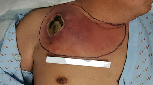
Right chest wall abscess with overlying eschar from extravasation injury due to calcium gluconate infusion via right internal jugular vein central catheter.
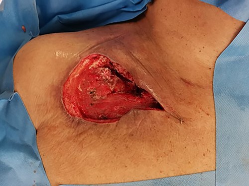
Intra-operative picture post second debridement.
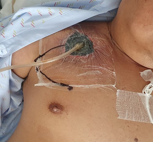
Post application of negative pressure dressing (after debridement).
29 days after the index surgery, he underwent a single stage ipsilateral pectoralis major turnover flap, rhomboid skin flap, and inferior nipple transposition (Figs. 4 and 5). A rhomboid skin flap based inferiorly was designed and raised. The right nipple was islanded on a central pedicle over the pectoralis major, allowing distal undermining without distortion of the nipple. The pectoralis major was incised over the nipple and turned up into the defect, preserving the internal mammary artery perforators and thoracoacromial pedicle. The defect was then obliterated and the pectoralis muscle flap was anchored. The skin defect was then closed with the rhomboid skin flap, and the nipple subsequently transposed inferiorly to match the contralateral side.
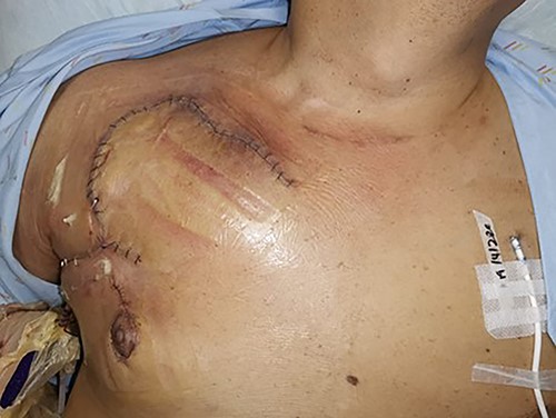
Post single stage right pectoralis major turnover flap, rhomboid skin flap, and inferior nipple transposition.
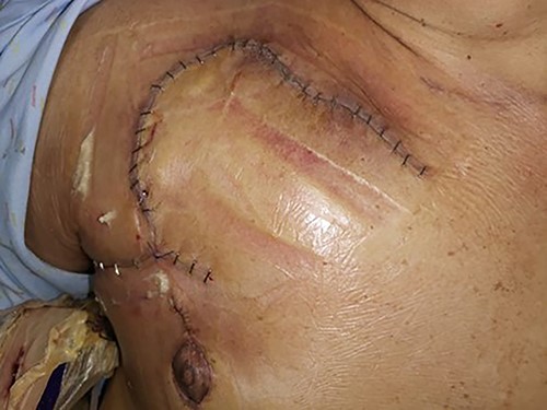
Close-up post single stage right pectoralis major turnover flap, rhomboid skin flap, and inferior nipple transposition.
Patient’s post-operative recovery was unremarkable except for a small haematoma over the medial aspect of the flap that resolved with conservative treatment, and he was discharged soon after. Reviews in clinic thereafter were unremarkable. He had no features of infection, no nipple necrosis, and regained full range of motion of his right upper limb. Patient was very satisfied with the end result, including nipple position (Figs. 6, 7, 8).
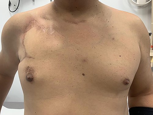
9 months post-operatively (anterior view).
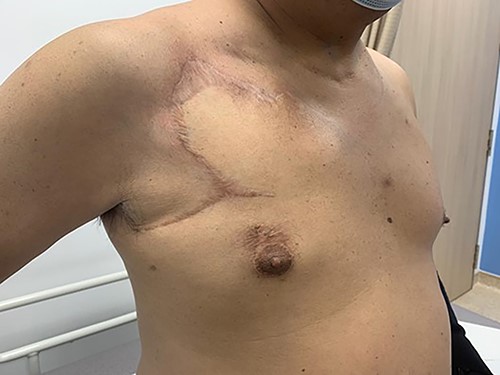
9 months post-operatively (anterolateral view).
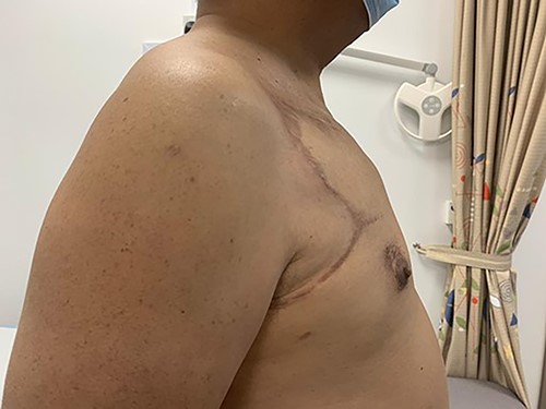
9 months post-operatively (right lateral view).
DISCUSSION
While chest wall soft tissue reconstruction has well been described – from pectoralis major flaps, to rectus abdominis flaps, to omental flaps – little has been described about the overlying skin reconstruction, as well as nipple positioning. To the best of our knowledge, this is the first description of a chest wall defect reconstruction with a single stage ipsilateral pectoralis major turnover flap, rhomboid skin flap, and inferior nipple transposition to match the contralateral side. This single stage surgery allowed the wound to be covered, as well as nipple to be matched to the contralateral side, in a single stage - avoiding future surgeries (and its associated risks). There was need to transpose the nipple inferiorly as insetting the inferiorly-based rhomboid skin flap over the wound would have resulted in a significantly higher right nipple if no transposition was performed. Keeping the nipple islanded on a central pedicle intra-operatively allowed for a dependable nipple vascular supply, and this likely contributed to the lack of nipple necrosis post-operatively.
Goals of this surgery included 1) coverage of wound with a well vascularised flap to obliterate dead space and combat infection, 2) regain right upper limb full range of motion, 3) symmetrical nipple placement bilaterally – and these were all achieved. This is promising as studies have described a 7% thirty-day perioperative mortality, and 80% complication rate (ranging from superficial wound dehiscence, seroma, to deep infection requiring further debridement) [2] for chest wall reconstructions. Pectoralis major flaps are also associated with decreased range of motion of the ipsilateral shoulder [5], for which this patient did not have. While no studies have described rates of nipple necrosis after chest wall soft tissue reconstruction, rates of nipple necrosis after nipple sparing mastectomies range up to 8% and 16% for total and partial necrosis respectively [6]. This patient had no nipple necrosis.
Regarding extravasation injuries, they are estimated at a rate of 0.1–6% [7], with serious extravasation injuries estimated at 0.01–0.1% [8]. Extravasation injuries via central catheters can progress to involve the mediastinum / pleural space, causing potentially life-threatening complications. As such, a CT thorax scan was done to ensure vital surrounding structures were not affected. With no vital structures seen affected in the CT thorax, debridement was carried out and reconstruction planned for the defect.
In conclusion, we propose that a single stage ipsilateral pectoralis major turnover flap, rhomboid skin flap, and inferior nipple transposition is a viable reconstructive option for anterior chest wall soft tissue defects.
DATA AVAILABILITY
The data of this case report are available from the corresponding author upon reasonable request.
CONFLICT OF INTEREST STATEMENT
None declared.
FUNDING
None.


