-
PDF
- Split View
-
Views
-
Cite
Cite
Isaac Ealing, Ross Calopedos, Marnique Basto, Catalina Palma, John Boulas, Anatrophic nephrolithotomy and pyelolithotomy for dual staghorn calculi in bifid collecting system, Journal of Surgical Case Reports, Volume 2022, Issue 11, November 2022, rjac547, https://doi.org/10.1093/jscr/rjac547
Close - Share Icon Share
Abstract
A bifid ureter is an atypical anatomical variation that occurs with an incidence of 1–10%. This anomaly is in a continuum of duplex collecting systems and most commonly involves a common distal ureter. This is usually asymptomatic and is predominantly an incidental diagnosis, nevertheless, is a potential risk factor for urolithiasis formation. Current surgical management of larger staghorn calculi favours percutaneous nephrolithotomy (PCNL) over traditional open surgery, however for multiple calculi and complex anatomy PCNL would require multiple punctures, with increased risk of bleeding, pleural injury, sepsis and ultimately failed stone clearance. We describe the case of a 71-year-old female with multiple calculi in bifid anatomy. A single open approach, aided with cold-ischaemia was successfully utilized in this context.
CASE REPORT
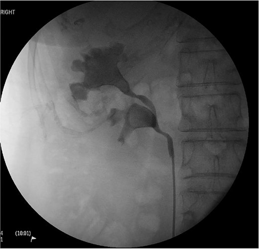
A 71-year-old female presented with urosepsis, right flank pain and a history of recurrent UTIs. CTKUB noting large right-sided staghorn calculi within the superior pole and renal pelvis without dilatation. Retrograde pyelogram further demonstrating bifid renal pelvis (Fig. 1). She was treated for Pseudomonas aeruginosa with IV piperacillin-tazobactam, and prepared for inpatient definitive management. Pre-operative creatinine was 65 umol/L, and haemoglobin 130 g/L.

Right retrograde pyelogram as part of pre-operative workup and diagnosis. Evidence of a proximal bifid ureter with distal confluence can be seen, and the presence of two large staghorn calculi are clearly seen in both the upper and lower moity renal pelvis.
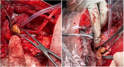
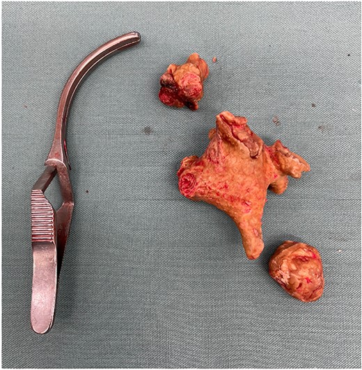
She underwent an open retroperitoneal lower moiety pyelolithotomy and upper pole anatrophic nephrolithotomy via supra-12th rib flank incision. After mobilization within Gerota’s fascia, the proximal renal artery and the posterior segmental branch were isolated. A longitudinal pyelotomy over the lower pole moiety was aided with use of eye-lid retractor sinus elevation as available ‘Gil Vernet’ retractor was too large. A 2-cm staghorn calculi was extracted (Figs 2 and 3). The longitudinal pyelotomy was closed with interrupted 3-0 chromic suture. Brodel’s line demarcation was then achieved by posterior segmental artery clamping and marked with diathermy. The main renal artery was clamped and ischaemic hypothermia maintained with ice-slush. A scalpel blade handle was used to fracture parenchyma until upper pole moiety entered. A 5-cm staghorn calculi was mobilized from adherent collecting system with aid of brain retractor and Randall’s forceps (Figs 2 and 3). Calicoplasty/caliorrhaphy was not required as all branches were easily accessible. Nephrotomy was closed with 2-0 chromic suture, incorporating urothelium, parenchyma and capsule. Suction drain was placed outside of re-approximated Gerota’s fascia and wound closed in layers. The patient’s recovery was uncomplicated and she was discharged day eight with creatinine of 41umol/L and haemoglobin 106 g/L.

(Left) Removal of the lower moity staghorn calculi via a pyelotomy at the base of the right inferior moity renal pelvis. (Right) Removal of the upper moity staghorn calculi via a posterior parenchymal incision along the bloodline plane of Brodel (between the anterior and posterior segmental branches of the renal artery.

Both the upper (with additional fragment above) and lower staghorn calculi specimen placed next to a bulldog vascular clamp for reference.
DISCUSSION
A bifid ureter is an atypical anatomical variation that occurs with an incidence of 1–10% [1–3]. This anomaly is in a continuum of duplex collecting systems and most commonly involves a common distal ureter. This is usually asymptomatic and is predominantly an incidental diagnosis, nevertheless, is a potential risk factor for urolithiasis formation [4–6]. Current surgical management of larger staghorn calculi favours percutaneous nephrolithotomy (PCNL) over traditional open surgery [7, 8]. However, with added complexity of multiple calculi in bifid anatomy, percutaneous approaches require multiple punctures and prolonged ureteroscopic laser lithotripsy [9, 10]. This increases bleeding risk, pleural injury, sepsis and ultimately failure stone clearance [9]. A single open approach, aided with cold-ischaemia should be considered in these cases. Open, anatrophic nephrolithotomy involves opening the renal pelvis from a posterior, intersegmental parenchymal incision along the bloodless plane of Brodel. This has been described for large staghorn calculi, often with complex anatomy in several case series [8, 10–13]. To the best of our knowledge anatrophic nephrolithotomy with pyelolithotomy has not been described in the literature for dual staghorn calculi in a bifid ureteric collecting system. In conclusion, the concurrent presence of large stone burden and complex anomalous anatomy require a comprehensive workup; understanding of the individual patient anatomy; and consideration of open surgical nephrolithotomy.
CONFLICT OF INTEREST STATEMENT
None declared.
FUNDING
None.


