


Abstract
Glomus tumor (GT) constitutes a rare, benign, soft-tissue tumor emerging from neuro-myo-arterial glomus bodies. Due to its rarity, and absence of typical symptoms, GT is usually misdiagnosed, with a potential risk of rupture and infection, or even malignant transformation. The present manuscript reports a rare case of a 17-year-old young woman with multiple GTs in her lower back, breach and left thigh that was surgically treated. The manuscript aims to highlight the importance of prompt diagnosis and surgical treatment of this peculiar tumor in young patients and raise surgeons’ awareness.
INTRODUCTION
Glomus tumor (GT) constitutes a rare, benign, soft-tissue tumor emerging from neuro-myo-arterial glomus bodies [1]. Although GT is mostly found in hands, it may develop anywhere in the human body and present with the classic triad of pain, hypersensitivity to cold and tenderness [2, 3]. Due to its rarity, and absence of typical symptoms, GT is usually misdiagnosed and subsequently it is not treated adequately [4]. However, such a malpractice is extremely severe, since GT involves the risk of rupture and infection, malignant transformation and metastases [1, 2]. The present manuscript reports a rare case of a 17-year-old young woman with multiple GTs in her lower back, breach and left thigh, and aims to highlight the importance of prompt diagnosis and surgical treatment of this peculiar tumor in young patients.
CASE PRESENTATION
A 17-year-old Caucasian female proceeded to our institution complaining of a painful, palpable nodule on her left buttock and thigh for the last 2 months. The patient mentioned that the pain was reduced by rest, but it was exacerbated in the sitting position and upon palpation. Motion and sensation over the leg were unaffected. Physical examination revealed a blue-red painful soft mass on her left buttock (Fig. 1). The overlying skin had no evidence of inflammation or trauma. A similar mass, with mild tenderness upon palpation was detected on her left thigh as well (Fig. 2).
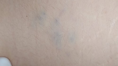
A blue-red painful soft mass on the patient’s left buttock.
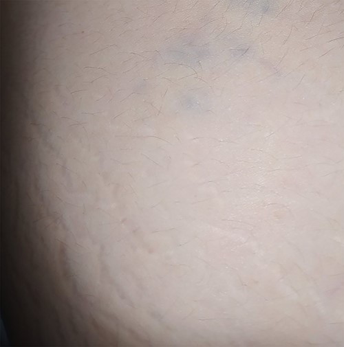
A similar blue-red painful soft mass was detected on her left thigh.
Laboratory examinations, including CRP and ESR where within the normal spectrum. The patient’s medical and surgical history was unremarkable. A subsequently ordered ultrasound detected an endogenic rounded soft-tissue mass with a diameter of ~3 cm on her left buttock, consisting with a hypervascular nerve tumor on Doppler examination. Similar findings were detected when investigating the mass over the patient’s left thigh. Based on the above findings, a surgical resection of the masses were scheduled.
During the procedure tumors were removed completely and subsequently ordered pathological examination confirmed the diagnosis of a GT (Figs 3–5). The report documented the presentation of benign tumors with dilated vessels, surrounded by collars of glomus cells with adjacent hyalinized stroma. The operation was uneventful and on a follow-up visit 6 months later, the patient was completely pain free, with no evidence of local recurrence (Fig. 6). However, a similar lesion was found on the left side of the patient’s lumbar region.
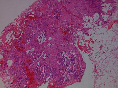
Hematoxylin and Eosin ×40: pieces of neoplasm with adjacent fatty tissue.
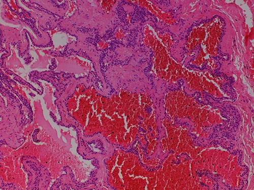
Hematoxylin and Eosin ×100: dilated vessels surrounded by collars of glomus cells with adjacent hyalinized stroma.
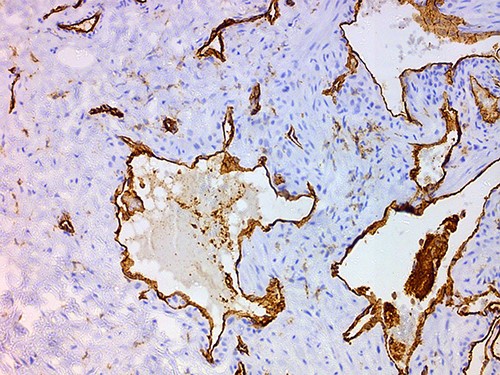
CD31 ×100: endothelial cells express CD31 whereas neoplastic surrounding cells do not.
Another similar mass found on the patient’s lower back.
DISCUSSION
GT is a rare hamartoma, arising from neuro-myo-arterial glomus bodies accounting for 1% of soft-tissue tumors [1–3]. GT was first described by Wood in 1812 as a painful blue-red tumor [1]. Although GT is typically presented in the subungual region of hands, and the lateral area of the fingers, it may develop anywhere at the dermo-epidermic junction [2]. Less frequently, GT may be detected on the leg, breast, toes or even the lungs [2, 5–7].
In the currently presented case, multiple GTs were detected in less common anatomical regions in a young female. GT may appear at any time during adulthood with a 4:1 male to female predominance [8]. Clinical presentation of GT typically includes tenderness upon palpation, intolerance to cold and pain [3, 8].
Diagnosis of GT before its resection is extremely difficult to reach to, because of its rarity and its vague clinical symptoms [1, 3]. Nevertheless, ultrasonography is extremely beneficial, since detecting a hypo-echoic, well-demarcated mass, which appears hyper-vascularized on Doppler, suggests the diagnosis [9, 10]. Computed tomography scan and angiography may be useful as well, but magnetic resonance imaging better delineates the margins of the tumor from the surrounding normal tissues [3, 9, 10]. However, the diagnosis is confirmed only by the histopathological examination, as in the currently presented case.
GT is mainly treated by surgical resection. The procedure is performed when the patient suffers from hypersensitivity and pain, or in cases of possible malignancy or infection [1, 2, 10]. In such cases, meticulous histological observation, and utilization of immunohistochemical markers is of paramount significance, to confirm the diagnosis of malignancy, elucidate the malignant potential and determine the appropriate therapeutic approach [2]. Finally, recurrence after surgical removal is potential, and therefore regular follow-up visits are of paramount clinical significance.
Conclusively, GT is a rare benign soft-tissue tumor that is usually misdiagnosed and inappropriately treated. Insufficient surgical treatment, without meticulous resection of the tumor, leads to unbearable pain, swelling, limited motion or even risk of malignant transformation. Hence, surgeons’ in-depth knowledge and high clinical suspicion is fundamental and crucial for the diagnosis, treatment and follow-up of GT.
CONFLICT OF INTEREST STATEMENT
None declared.
