-
PDF
- Split View
-
Views
-
Cite
Cite
Dafina Mahmutaj, Bedri Braha, Jehona Krasniqi, Gastro-cutaneous fistula treated with trionic (cell alginate activate packing), Journal of Surgical Case Reports, Volume 2021, Issue 8, August 2021, rjab346, https://doi.org/10.1093/jscr/rjab346
Close - Share Icon Share
Abstract
Gastrocutaneous fistulae are traditionally treated with to parenteral nutrition or surgical management. We are presenting a case of a 56-year-old man who underwent a surgical closure of a gastrocutaneous fistula with a trionic (cell alginate activate packing). The fistula recurred on postoperative day 14, after the Bilroth II operation.
For the first 8 days after filling the fistula with trionic, we applied total parenteral nutrition. Later, the patient started taking liquid foods through the mouth. The leak of the fistulous liquid was conspicuously reduced, and on the 20th day, it ceased completely.
INTRODUCTION
Gastrocutaneous fistula results from abnormal communication between the stomach and the skin. It has an inner hole, an outer hole and a canal that is filled with epithelial cells. Gastrocutaneous fistula is a rare complication of the upper gastrointestinal tract surgery, occurring only in 0.5–3.9% of cases. Nevertheless, it has high mortality and considerable morbidity when manifested [1]. Gastrocutaneous fistulas close spontaneously only in 6% of the cases, and in the remainder of patients (with a normal weight) that undergo surgical intervention in the stomach, the mortality rate is ~35% [2].
Gastrocutaneous fistula occurs in: dehiscence of gastroenteral anastomosis, disruption of stomach after resection, iatrogenic injuries and failure of resolution of fistula after the removal of the gastrostomy tube. The pathogenesis implied in iatrogenic injuries of the stomach and disruption of sutures placed in the stomach has a root cause in vascular necrosis [3, 4]. An important element in the formation of gastrocutaneous fistulas is the epithelialization of the fistula tract. Based on this, chronic gastrostomies (>6 months) can epithelialize and become persistent fistulas [5]. The application of radiotherapy effects the gastric mucosa, and it provokes the recurrence of gastric ulcers. These ulcers can perforate and form abscess and fistula [6].
CASE PRESENTATION
We present a 56-year-old man, admitted at the Surgery Clinic with the diagnosis of stomach carcinoma localized at the pre-pyloric region of the stomach. After proper preparation, the patient went through the surgery. The operative procedure that was performed was a partial gastrectomy and gastrojejunostomy sec Bilroth II with lymphadenectomy. Owing to the transverse colon obstruction and infiltration, we performed a resection with termino-terminal anastomosis.
The postoperative state of the patient was stable with normal radiologic and lab results. The histology report after the operation was: stomach adenocarcinoma (intestinal type, Gr. II, pT3, pN0).
After 14 days, the patient had begun to complain of abdominal pain. The laboratory test results showed an increased white blood cells, with an increase in the biomarkers of infection (C-reactive protein and procalcitonin). The wound had started leaking considerable purulent discharge with a foul smell. A swab was taken from the operating wound where Pseudomonas aeruginosa was isolated. The empiric therapy had been initiated until microbiological analysis, after which we continued the treatment according to the antibiogram. A colostomy bag was placed at the site of the leak and the amount of the contents was measured. The amount was about a 1000 ml within 24 h, and the contents smelled like vomit. The contents in the colostomy sac were gastric contents, gauging from the physical properties. This leak continued on the second day with the same amount of the content. Therefore, the patient began to be treated with total parenteral nutrition. We tested if the fistula was leaking by giving the patient a red liquid to drink. We noticed that the liquid was leaking out of the fistula. The overall health of the patient was bad. After a proper resuscitation through consultation with other doctors of the department, we decided to fill the fistula with a trionic material. The filling of the fistula with trionic material was done under general anesthesia, as shown in Fig. 1.
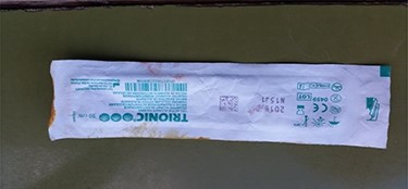
Several steps were taken during the procedure of the filling of the gastrocutaneous fistula under general anesthesia:
Step 1: Cleaning of the operating field with iodine. Because of the non-formation of the outer opening of the fistula in the skin, the skin was opened. A foley catheter was placed in the fistula with the purpose of stopping the leakage, and was evaluated, as shown in Fig. 2.
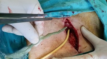
Identification of gastrocutaneous fistula and placement of foley catheter. Gradual retraction of the foley catheter and preparation of the filling by twisting it and holding it in the right direction (the foley catheter as a guide).
Step 2: The operating field was cleansed, after which the filling was put inside the fistula by twisting it gently as shown in Fig. 3.
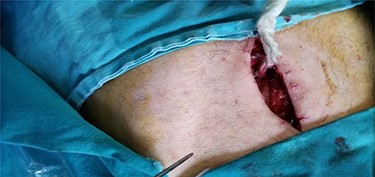
Filling is placed and fixed to the wall around the external fistula orifice and to the fascia.
Step 3: Being careful not to move the filling, two sutures were put in place adjacent to the wall of the fistula canal identified earlier, and two other sutures were inserted in the fascia, as shown in Fig. 3.
Step 4: The skin was stitched close, and the filling was fixed to the skin, as shown in Fig. 4.
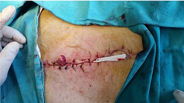
Trionic filling fixed with two sutures for skin and skin closure.
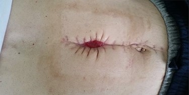
After the intervention, for the first 8 days, the patient was put on total parenteral nutrition. On the 9th day, the patient started consuming small amounts of liquid food. In the subsequent days, the amount of the food has gradually increased. In the place of the filling, a colostomy sack was inserted to measure the amount of leakage, which for the next 24 h was 5–20 ml. The leakage had considerably reduced until the 20th day when it finally stopped, as shown in Fig. 5. The operated wound had been cleansed every day, mainly with saline solution. The closure of the wound was per secundam intentionem. After the fistula was healed, the patient continued the treatment in oncology.
Trionic (cell alginate activate packing)
The trionic solution is composed of brown algae, chlorophyll, calcium, zinc and magnesium ions, which help regenerate tissues. Trionic absorbs the purulent discharge and it limits the spread of the necrotic tissue and bacterial invasion. In contact with natural secretions of the body, ringer solution, saline solution 0.9%, the trionic solution forms a gelatinous mass, which forms a hospitable environment that promotes wound healing.
DISCUSSION
The utilization of filling substances to fill anal fistula during the gastrocutaneous fistulas is rare, and mainly combined with the closure of the inner opening of the fistula with an endoscope, in which case a clip is put in place.
Darrien and Kasem have a series of seven patients whose fistula had been filled with Surgisis, two cases in which besides filling there had also been a clipping of the inner opening with an endoscope, whereas in other cases only the filling had been done. In these seven cases, there were no further problems [7]. Wood, with his fellow researchers, demonstrated a successful closure of a persistent gastrocutaneous fistula after removal of feeding gastrostomy in combination of clip in the inner opening with endoscopy [8].
Other alternative therapies for treating gastrocutaneous fistula include using a filling of Vicryl and fibrous glue [9]. There are some surgeons who apply over the scope clips. Lukish has used 2-octylcyanoacryatin for filling for gastrocutaneous fistula [10].
We have not come across any cases of filling of the gastrocutaneous fistula with trionic in existing literature.
COMMENTS
Such a method is minimally invasive and a good option in treating gastrocutaneous fistula, especially if patients are not qualified for surgical treatment because of their overall bad health. Nevertheless, it is imperative that a higher number of patients are treated, so that we can come to definitive conclusions about whether this method has more to offer.
CONFLICT OF INTEREST STATEMENT
None declared.


