-
PDF
- Split View
-
Views
-
Cite
Cite
João L Pinheiro, Marisa Marques, Carlos Daniel, Jorge Pereira, Carlos Casimiro, Duodenal recurrence of endometrial carcinoma: report of a rare metastatic site, Journal of Surgical Case Reports, Volume 2021, Issue 5, May 2021, rjab209, https://doi.org/10.1093/jscr/rjab209
Close - Share Icon Share
Abstract
Endometrial carcinoma is one of the most common gynaecologic malignancies in the western society. Treatment of recurrent disease became more refined, with the study of molecular and hormonal receptors playing a central role. A 76-year-old caucasian woman presented to the emergency department with growing tiredness, and melaena. Past medical history included an endometrioid adenocarcinoma. The patient had undergone a hysterectomy with bilateral salpingo-oophorectomy with pelvic and paraaortic lymphadenectomy and was disease-free for 2 years. The endoscopy revealed an ulcerated lesion involving the second and third portions of the duodenum. Histopathologic examination confirmed a poorly differentiated adenocarcinoma of endometrial origin. She started palliative chemotherapy, remaining with adequate symptomatic control. Endometrial cancer recurrence typically occurs locally. The liver is the intra-abdominal organ most commonly involved. There are scarce reports of duodenal metastasis of malignancies originated in distant organs. The duodenum remains an uncommon metastization site and is rarely associated with endometrial cancer.
INTRODUCTION
Endometrial cancer is a growing concern in the western society. Its incidence is rapidly rising along with obesity, the most important risk factor for this disease. When not associated with Lynch syndrome, the average age of diagnosis lies in the sixth decade of life [1–4]. Currently, molecular and hormonal characterization is of the utmost importance when treating advanced disease, allowing for targeted systemic therapy and increased overall survival [1, 3]. Around 64% of recurrences occur within 2 years after the first treatment. Frequent sites of disseminated endometrial carcinoma include pelvic and para-aortic lymph nodes, vagina, peritoneum and lungs. Less commonly, there have been reports of metastases found in bones and in the central nervous system [3, 5]. When extrapelvic spread occurs, endometrial cancer is considered incurable and all therapeutic approaches are of palliative intent [6]. For such patients, a platinum-based chemotherapy regimen is recommended [1, 4].
CASE REPORT
A 76-year-old caucasian woman presented to the emergency department (ED) with growing tiredness, melaena and left upper quadrant discomfort that worsened in the previous 10 days. Past medical history included a thyroidectomy due to multinodular goiter, and a hysterectomy with bilateral salpingo-oophorectomy with pelvic and paraaortic lymphadenectomy due to an endometrioid adenocarcinoma (pT1aN0M0, G3), which was diagnosed 2 years prior to the patient attending the ED. A follow-up computed tomography (CT) had ruled out any secondary lesions and tumour markers remained within normal limits. The patient was then started on vaginal brachytherapy and for 2 years had no recurrence documented.
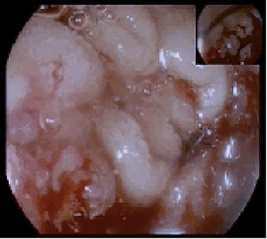
The patient was also being studied in our outpatient clinic for a symptomatic iron deficiency anaemia and had an upper gastrointestinal (GI) endoscopy performed the day before attending the ED. The endoscopy revealed a proliferative and ulcerated lesion involving the transition between the second and third portions of the duodenum (D2–D3; Fig. 1).

Upper endoscopy showing an ulcerative and friable duodenal lesion.
Due to the haemorrhagic risk, no biopsies were taken. She had no prior complaints of weight loss, nausea or vomiting. The full blood count revealed a haemoglobin of 7.5 g/dl and an haematocrit of 24%. She remained haemodynamically stable and, after optimization with a transfusion of 2 units of packed red blood cells, a second endoscopy and a colonoscopy were performed to obtain biopsies and exclude other causes of GI bleeding. She was admitted to the ward for further investigation.
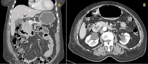
During the hospital stay, an imaging workup was carried out while awaiting the histological diagnosis and the patient had a thoracic, abdominal and pelvic CT done (Fig. 2A and B).

(A) Coronal cut of the abdomen on CT showing the third portion of the duodenum visibly thickened (arrow); (B) Arterial phase showing the circumferential involvement of D3 (arrow).
It described an almost circumferential thickening of the second and third duodenal portions consistent with the lesion described in the endoscopy, extended for 4 cm, with 12 mm of width, and several perilesional adenopathies, the largest measuring 9 mm.
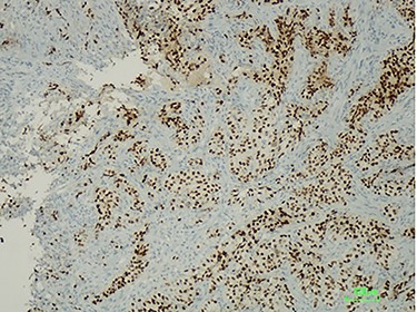
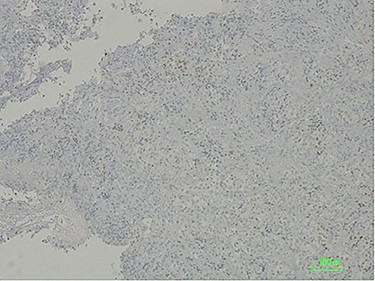
The histologic examination revealed a poorly differentiated adenocarcinoma, with a negative CDX2 and positive PAX8 immunostaining, hence compatible with a secondary lesion of endometrial origin (Figs 3 and 4).

Immunostaining positive for PAX8 suggesting a tumour of gynaecologic origin based on the patient’s past medical history.

CDX2 negative immunostaining excluding carcinoma of intestinal origin such as primary duodenal neoplastic lesion.
The case was discussed in a multidisciplinary meeting and the patient started palliative chemotherapy with carboplatin and paclitaxel, remaining to this day with adequate symptomatic control.
DISCUSSION
Endometrial carcinoma is one of the most common gynaecologic malignancies in the western society [2, 4]. The incidence of this type of cancer has been increasing along with the higher rates of obesity, its major risk factor [1]. In the past decade, the surgical treatment has evolved with the addition of sentinel node mapping. As for the treatment of recurrent disease it became more refined, with the study of molecular and hormonal receptors having a central role when choosing the adequate systemic treatment [1, 4].
After surgical resection, the patient was initially staged as a Federation of Gynaecology and Obstetrics IA Grade 3 pT1aN0M0 endometrioid adenocarcinoma, the most common histologic type of endometrial cancer, accounting for 80% of all endometrial carcinomas. The neoplastic cells of the resected specimen were negative for estrogen and progesterone receptors and there was a loss of expression of the MLH1 and PMS2 proteins which, given the age of presentation and lack of familiar history, were in accordance with a sporadic cancer [1, 4]. The duodenal lesion was not tested for hormonal receptors as the primary tumour was already known to be negative for both.
Endometrial cancer recurrence is typically local, either involving the vagina, or pelvic and para-aortic nodes, commonly seen in patients treated with chemotherapy alone [2, 5]. Extra-nodal involvement is uncommon at the time of diagnosis. Peritoneal carcinomatosis is found in ~28% of relapses from simple serosal implants, to extensive peritoneal masses that can present as intestinal obstruction by extrinsic compression.
Recurrence in intra-abdominal organs is rare and often associated with aggressive histopathologic types such as the clear cell and serous type. Although uncommon, endometrial cancer has the highest incidence of lung dissemination of all gynaecologic cancers, going up to 25%. The liver is the most commonly involved intra-abdominal organ, with secondary lesions in 7% of cases [6, 4]. Muscular and soft tissue recurrences are rare and account for 6% of all distant metastatic sites. Other atypical secondary lesion locations include the bone, adrenal glands and the spleen. In less than 1% of cases, there is central nervous system involvement [5]. The metastatic dissemination pathway to the duodenum remains unclear. In such cases, retrograde lymphatic spread from the para-aortic lymph nodes is a possible mechanism [7].
To this day, despite direct invasion being quite common, there have been very few reports of duodenal metastasis of malignancies originated in distant organs. Lung, renal, melanoma and colorectal cancer have sporadic reports of duodenal involvement [8, 9]. When identified on endoscopy, they frequently present as ulcerated lesions that can cause GI bleeding or gastric outlet obstruction. This finding is consistent with the endoscopy’s macroscopic description of the duodenal lesion and our patient’s clinical presentation. Immunohistochemistry provided the diagnosis after confirming its non-lower GI tract origin and gynaecologic related epithelial structure (positive for cytokeratin 7, negative CDX2 and positive PAX8 immunostaining) [3, 10].
The duodenum remains an uncommon metastization site for any distant primary tumour and is rarely associated with gynaecologic cancers such as endometrial adenocarcinoma.
CONFLICT OF INTEREST STATEMENT
The authors have no conflicts of interest to report.
FUNDING
The authors report no financial support.
References
- cancer
- ulcer
- adenocarcinoma
- endometrial cancer
- endoscopy
- fatigue
- carcinoma, endometrioid
- emergency service, hospital
- hysterectomy
- lymph node excision
- melena
- neoplasm metastasis
- abdomen
- duodenum
- medical history
- liver
- pelvis
- lymph node dissection
- endometrial carcinoma
- salpingo-oophorectomy
- gynecologic cancer
- play behavior
- palliative chemotherapy
- histopathology tests


