


Abstract
Dentigerous cyst is a type of developmental odontogenic cysts that arises from the crown impacted, embedded or unerupted teeth that standardly managed by Caldwell-luc procedure, which is found to be associated with morbidities and complications. Endonasal endoscopic removal is a minimally invasive approach aiming to prevent morbidities and complications. The aim of this article is to study the advantages of the endonasal endoscopic approach in managing different cases of dentigerous cyst and ectopic teeth. In this article, we reported three different cases (two pediatric and one adult), one presenting with unilateral dentigerous cyst with traumatic ectopic teeth in the maxillary sinus, and one presenting with bilateral dentigerous cysts and ectopic teeth in the maxillary sinuses, the third case of unilateral ectopic intranasal canine tooth. All managed by endonasal endoscopic approach, with no complications and complete recovery. The endonasal endoscopic approach is a minimally invasive surgical approach. With the use of different angled endoscopes and instruments, this approach is preserving physiological function while minimizing morbidity and preventing complications.
INTRODUCTION
Dentigerous cyst is a type of developmental odontogenic cysts [1]. It accounts for 20–24% of all jaw cysts [2, 3]. Dentigerous cyst arises from the crowns of impacted, embedded or unerupted teeth [2]. It commonly involves the mandibular third molar teeth [4]. Ectopic eruption of teeth is rare and if occurs, it commonly involves the dental arch, palate, and nose and rarely the maxillary sinus [3]. Only few cases of dentigerous cysts associated with ectopic teeth within the maxillary sinus has been reported in the literatures [4].
Dentigerous cyst and the ectopic teeth associated with it are standardly removed via a Caldwell-Luc procedure [5]. Although it gives direct visualization to the maxillary sinus, it is associated with more complications and morbidities [6]. Endonasal endoscopic removal is another approach used for the treatment of dentigerous cyst and ectopic teeth [6]. It is a minimally invasive approach that is believed to be associated with less morbidities and complications and more preservation of physiological function [6].
In this article, we aim to study the advantages of the endonasal endoscopic approach in managing different cases of dentigerous cyst and ectopic teeth.
CASE SERIES
Case 1
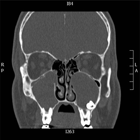
A 13-years-old boy medically and surgically free presented to the otolaryngology clinic with swelling of the right side of the face for 1 month, no other significant symptoms. Patient has history of facial trauma 8 years back. Examination was unremarkable apart from right-sided facial swelling over the maxillary sinus. Computed tomography (CT) of the paranasal sinuses showed a large expansible cystic lesion in the right maxillary bone that arises from the maxillary alveolar ridge with superior extension and displacement of the right maxillary sinus. A displaced tooth is seen within the anterior aspect with its root pointing posteriorly to the floor of the hypoplastic maxillary sinus with its crown embedded within the lesion (Fig. 1).
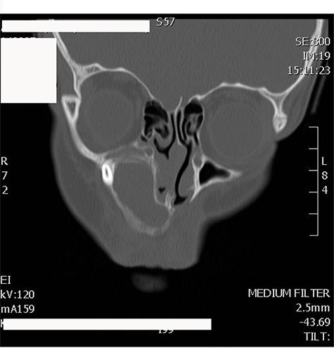
CT scan of maxillofacial bones showing a large expansible cystic lesion in the right maxillary bone that arises from the maxillary alveolar ridge with superior extension and displacement of the right maxillary sinus as well as remodeling of the floor of the right orbit with slight narrowing of the right inferior orbital fissure.
Informed consent was obtained from the parents and the patient underwent endoscopic endonasal enucleation of cyst and removal of ectopic tooth through inferior meatal antrostomy (Figs 2 and 3). Histopathologic examination confirmed the diagnosis of dentigerous cysts.
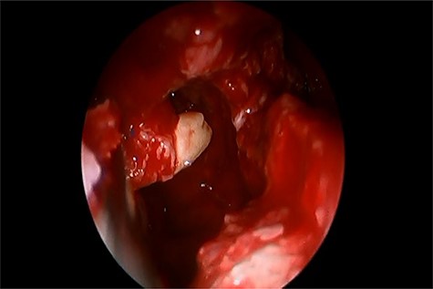
Inferior meatal antrostomy, after sac enucleation, while removing the tooth.
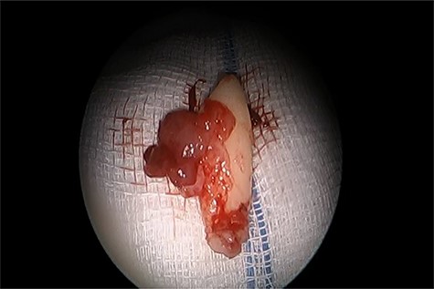
The ectopic tooth after removal.
Case 2
An 11-years-old girl known case of acute lymphocytic leukemia on remission, presented to otolaryngology clinic with right-sided nasal obstruction along with occasional headache. Symptoms are progressive over a period of 1 year. No history of trauma, surgery or foreign body. Examination showed a collapsed right lower lateral cartilage, supratip depression and hard bony mass filling the right nasal cavity. No other remarkable findings. CT of the paranasal sinuses is done and it shows displaced right upper maxillary tooth with crown oriented inferiorly and medially toward and within the lower right anterior nasal cavity with no destruction of the adjacent structures (Fig. 4A and B). Patient was managed with anterior rhinoscopy and endoscopic-guided removal of the ectopic tooth without any complications. The ectopic tooth is found to be canine tooth (Fig. 4). Histopathologic report confirms the diagnosis of ectopic tooth. Post operatively, the patient symptoms improved completely, and she remained symptoms free for 18 months.
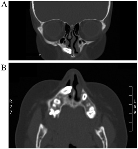
(A, B) CT of the paranasal sinuses (coronal and axial view) revealing displaced right upper maxillary tooth with the crown-oriented inferiorly and medially toward and within the lower right anterior nasal cavity.
Case 3
A 19-years-old male presented to otolaryngology clinic facial pain over the upper jaw area along with post-nasal discharge for 6 months. Patient gave history of recurrent sinusitis; otherwise, he is medically and surgically free with no history of trauma. Examination showed septal spur to the left side with no other significant findings. CT scan of the paranasal sinuses showed bilateral ectopic teeth and cystic lesions within both of the maxillary sinuses (Fig. 5). Patient was managed with endoscopic endonasal enucleation of the cysts and extraction of the ectopic impacted teeth through middle meatal antrostomies. Histopathology confirms the diagnosis of dentigerous cysts. Post operatively, the patient symptoms resolve completely, and she remained symptoms free over 5 years follow up.
CT scan of the paranasal sinuses showed bilateral ectopic teeth and cystic lesions within both of the maxillary sinuses.
DISCUSSION
Developmental cysts of the jaw are classified into an odontogenic group, arising from odontogenic tissue such as malassez epithelial remnants, dental lamina or enamel organ remnants and a non-odontogenic group, arising from ectoderm involved in the development of facial tissues [5, 7]. Dentigerous cysts are the second most prevalent type of odontogenic cysts after periapical or radicular cysts [5]. In 70% of the cases, they develop in the mandible, only about 30% of the cases occur in the maxilla [5]. They usually present in the second or third decade of life with male predisposition [5].
Dentigerous cysts are generally symptom free and many go unnoticed for several years. In such cases, they may be discovered incidentally on routine radiographic examination [5]. Symptoms of dentigerous cysts that invade the maxillary sinus may include facial pain, headache, purulent nasal discharge or nasolacrimal obstruction [5].
The standard treatment for a dentigerous cyst in the maxillary sinus is enucleation and extraction of the impacted tooth via combined endoscopic and Caldwell-Luc procedure [5]. There are multiple factors that influence the decision of surgical approach, such as extent of cyst invasion of surrounding structures, functional and cosmetic significance of the impacted tooth, cyst size and patient age [1]. If the cyst is small and in adult patient, eruption of ectopic tooth is impossible or a little chance, enucleation is preferred [5]. Marsupialization on the other hand, is recommended for large cysts to reduce the size of the osseous defect before going through enucleation and extraction of the tooth [5].
The endonasal endoscopic management of odontogenic cysts of the maxilla, including dentigerous cysts has been repeatedly described over the past 10 years in a total of 52 patients [7]. There were no reported recurrences when using these approaches and no major complications were associated with these procedures [7].
Different applications of the endoscopic technique for the treatment of dentigerous cysts, with or without associated ectopic teeth have been described in the literature.
Guang-zhou Xu et al. [8] performed a modified Caldwell-Luc approach for the treatment of dentigerous cyst, in which the bony lid had been anchored back in its original place.
Emanuelli et al. [6] proposed the endoscopic technique for the removal of ectopic tooth that is associated with a dentigerous cyst through combined lower and middle meatotomies.
Ma et al. [9] had two cases of maxillary dentigerous cysts with oroantral fistulas that been managed via an extended inferior meatal maxillary antrostomy through inferior turbinate.
Song et al. [10] described another endoscopic approach through anterior or posterior nasolacrimal duct for the treatment of maxillary sinus lesions.
Recently, endonasal endoscopic removal of large dentigerous cysts of the maxillary sinus has been also reported using the endoscopic approach through the middle meatus [7].
Although the traditional Caldwell-Luc procedure provides a direct view into the maxillary sinus, it is associated with more morbidities than is endonasal endoscopy [5]. It requires a large opening in the anterior maxillary sinus wall to extract the tooth [5]. This approach have carried a risk of complications, including damage to the sinus mucosa, retraction of the soft tissues of the cheek, oroantral fistula, infra-orbital nerve injury, and damage to the nasolacrimal duct and adjacent teeth [5].
The endonasal endoscopic approach by using the use of different angled endoscopes and instruments is a minimally invasive surgery preserving physiological function while minimizing morbidity and preventing complications [5].
Tooth and cyst that are close to the osteomeatel complex can be managed through a fully endonasal endoscopic approach, preferably through a middle meatal antrostomy. In our opinion, an exclusive endoscopic approach has to be considered even if the tooth is too far from the lateral nasal wall, or too close to the orbital floor or nasolacrimal duct, or if the cyst and tooth are too large.
CONCLUSION
This paper presented our experience in the management of different cases of dentigerous cysts and ectopic tooth using the endonasal endoscopic approach and avoiding the external approach with its complications and morbidities. The endonasal endoscopic approach by using the use different angled endoscopes and instruments is a minimally invasive surgical approach preserving physiological function while minimizing morbidity and preventing complications.
REFERENCES
Girish G, MaheshKumar R., DN U, Sharma R, Matamari V, Bhandari A. Dentigerous cyst in maxillary sinus: A rare occurrence.
