-
PDF
- Split View
-
Views
-
Cite
Cite
Danah I Alnahari, Sami A Almalki, Fahad A Alabeidi, Recurrent intestinal obstruction due to gallstone ileus during same hospital admission: a case report and literature review, Journal of Surgical Case Reports, Volume 2021, Issue 4, April 2021, rjab024, https://doi.org/10.1093/jscr/rjab024
Close - Share Icon Share
Abstract
Gallstone ileus (GSI) is an uncommon entity that causes obstruction of the intestinal lumen due to gallstones. It affects mainly the elderly with multiple comorbidities, leading to a high mortality rate. In this case, an 81-year-old woman was admitted due to GSI. She had a recurrence after 5 days of the index surgery. Recurrent intestinal obstruction due to GSI during the same hospitalization despite complete clearance of the small bowel from stones is rare. Through our case, we will discuss management along with a review of the current evidence.
INTRODUCTION
Gallstone ileus (GSI) is an uncommon entity that occurs when gallstones migrate to the bowel through a cholecysto-enteric fistula, causing mechanical small bowel obstruction. It presents clinically as mechanical small bowel obstruction in a patient with a background of long-standing gallstone disease or symptoms suggestive of it. Features of GSI in abdominal radiographs include pneumobilia, intestinal obstruction and the presence of an aberrant gallstone (Rigler’s triad) [1]. We report the case of an 81-year-old female with recurrent GSI during the same admission. Clinical presentation, investigations and management are discussed along with review of the current evidence
CASE PRESENTATION
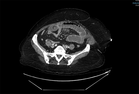
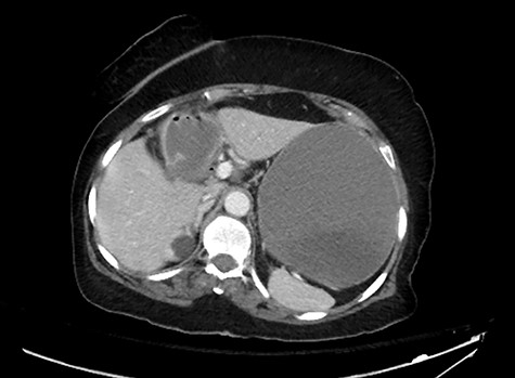
An 81-year-old woman with long history of gallstones was presented to the emergency department with persistent vomiting associated with progressive abdominal distension and inability to pass bowel motion or gases for the past 3 days. Examination showed stable vitals and distended abdomen. A computerized tomography (CT) scan showed high-grade small bowel obstruction in mid-ileum caused by a large impacted gallstone (Fig. 1). There was also a wide neck cholecystoduodenal fistula (Fig. 2).

A CT abdomen and pelvis scan showing a large stone in the small bowel.


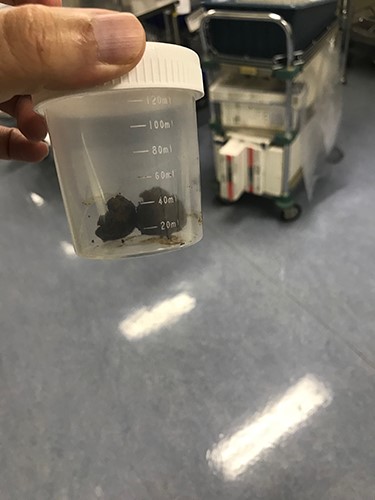
The extracted stone from the small bowel. Note that it was extracted as one piece.

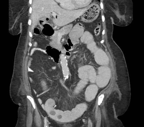
A CT abdomen and pelvis scan showing multiple large stones in the small bowel.
The patient was taken to the operating room (OR) for diagnostic laparoscopy that revealed small bowel obstruction with a transition zone at the mid-ileum. An enterotomy proximal to the obstruction point was made and the stone was removed. The stone was round in shape, not faceted (Fig. 3). The rest of the bowel was examined from the Ligament of Treitz to the ileocecal valve with no other stones identified. No attempt was made to explore the right upper quadrant as there was no plan to take down the fistula during this surgery.
Postoperative course was marked by a slow recovery evidences by persistently high nasogastric tube output and failure to open her bowel. On the third postoperative day, a trial of therapeutic Gastrografin was given, not much improvement was achieved, and the patient continued to be obstructed. A repeat abdominal CT scan was obtained on day 5 due to persistence of intestinal obstruction. It showed dilated contrast-filled small bowel loops with multiple filling defects, indicating recurrent GSI (Fig. 4).
The patient was taken back to the OR for diagnostic laparoscopy. Small bowel was markedly dilated and edematous, which made manipulation of small bowel loops difficult. Ascertaining locations of stones was not easy; therefore, laparoscopy was aborted and a mini-midline laparotomy incision was created. Palpation revealed multiple stones along the length of the small bowel. The previous enterotomy site was re-opened and the bowel was milked from the ligament of Treitz downward. A total of eight varying size stones were retrieved; largest measured 2 × 3 cm in size. Enterotomy site was used to perform a side-to-side stapled anastomosis. Postoperatively, the patient opened her bowels and tolerated diet. She left the hospital on the third postoperative day.
DISCUSSION
GSI is defined as an obstruction of the small bowel caused by gallstone impaction within the lumen of the gastrointestinal tract. GSI accounts for 5.3% of all cases of small bowel obstruction [2]. This percentage increases up to 25% in elderly patients with multiple comorbidities, which accounts for a higher rate of mortality ranging from 12 to 17% [3].
Patients usually present with non-specific signs and symptoms, leading to delay in diagnosis. Symptoms of small bowel obstruction including nausea, vomiting and crampy abdominal pain predominate. Plain abdominal images may aid in reaching the diagnosis by showing pneumobilia. Plain radiographs, however, have poor sensitivity ranging from 40 to 70% [4]. The widespread availability of CT scanning improved the diagnostic tools with a sensitivity of 93% [5].
Once impacted, spontaneous passage of these large stones is unlikely. Therefore, surgery is required for almost all symptomatic patients with GSI. An important clinical question lies in whether to deal with the biliary pathology at the index operation by closing the fistula and performing cholecystectomy or to simply address the obstruction by removing the gallstone with enterolithotomy. In one study, the mortality rate was higher (16.7%) for patients undergoing enterolithotomy along with addressing the biliary pathology in the same operation compared with enterolithotomy only (11.7%) [6].
The risk of GSI recurrence ranges from 5 to 8% [6–8]. However, these numbers are based on published case reports or series, which can underestimate the actual percentage. Approximately 50% of recurrent cases occur within the first month [9]. Of note, patients requiring surgery due to recurrence have higher mortality rates ranging from 12 to 20% [6, 10].
In the case described, the patient did not recover after the index surgery with recurrent GSI in the same admission. To the best of our knowledge, this case has the shortest recorded time for recurrence. Of note, a meticulous search for additional stones was carried out during index surgery and the initial CT scan was reviewed again retrospectively for additional stones outside of the gallbladder other than the one causing obstruction, and there were none. Recurrence could be attributed to the large fistula size, which allowed more stones to pass.
CONCLUSION
GSI is a true mechanical obstruction affecting mainly elderly patients. Surgery is required to address the obstruction. Current management is mainly based on published case reports or series with small numbers of patients. Enterolithotomy alone seems to be the safest surgical intervention. Recurrent GSI carries a higher risk of morbidity and mortality.
CONFLICT OF INTEREST STATEMENT
None declared.
FUNDING
None.
CONSENT
The patient has consented to this publication. All measures were taken to ensure anonymity and privacy.


